. 2025 Jan;21(1):160-177.
doi: 10.1080/15548627.2024.2394306. Epub 2024 Sep 10.

Phosphorylation of the selective autophagy receptor TAX1BP1 by TBK1 and IKBKE/IKKi promotes ATG8-family protein-dependent clearance of MAVS aggregates

Affiliations

Phosphorylation of the selective autophagy receptor TAX1BP1 by TBK1 and IKBKE/IKKi promotes ATG8-family protein-dependent clearance of MAVS aggregates

Jesse White et al. Autophagy. 2025 Jan.

Abstract

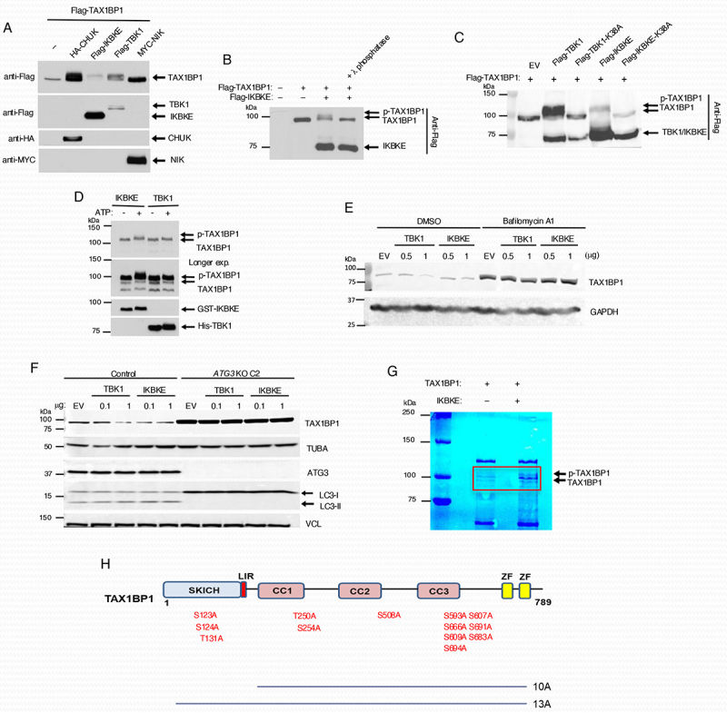
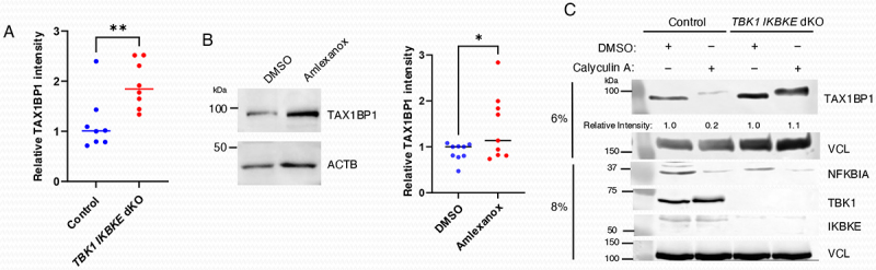
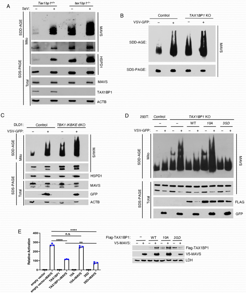
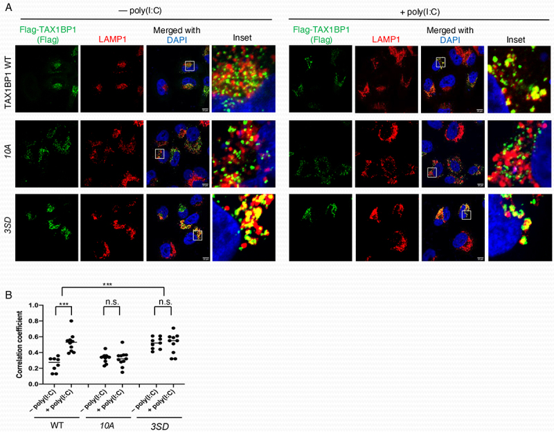
TAX1BP1 is a selective macroautophagy/autophagy receptor that inhibits NFKB and RIGI-like receptor (RLR) signaling to prevent excessive inflammation and maintain homeostasis. Selective autophagy receptors such as SQSTM1/p62 and OPTN are phosphorylated by the kinase TBK1 to stimulate their selective autophagy function. However, it is unknown if TAX1BP1 is regulated by TBK1 or other kinases under basal conditions or during RNA virus infection. Here, we found that TBK1 and IKBKE/IKKi function redundantly to phosphorylate TAX1BP1 and regulate its autophagic turnover through canonical macroautophagy. TAX1BP1 phosphorylation promotes its localization to lysosomes, resulting in its degradation. Additionally, we found that during vesicular stomatitis virus infection, TAX1BP1 is targeted to lysosomes in an ATG8-family protein-independent manner. Furthermore, TAX1BP1 plays a critical role in the clearance of MAVS aggregates, and phosphorylation of TAX1BP1 controls its MAVS aggrephagy function. Together, our data support a model whereby TBK1 and IKBKE license TAX1BP1-selective autophagy function to inhibit MAVS and RLR signaling.Abbreviations: ATG: autophagy related; BafA1: bafilomycin A1; CALCOCO2: calcium binding and coiled-coil domain 2; GFP: green fluorescent protein; IFA: indirect immunofluorescence assay; IFN: interferon; IκB: inhibitor of nuclear factor kappa B; IKK: IκB kinase; IRF: interferon regulatory factor; KO: knockout; LAMP1: lysosomal associated membrane protein 1; LIR: LC3-interacting region; MAP1LC3/LC3: microtubule associated protein 1 light chain 3; MAVS: mitochondrial antiviral signaling protein; MEF: mouse embryonic fibroblast; MOI: multiplicity of infection; IKBKG/NEMO: inhibitor of nuclear factor kappa B kinase regulatory subunit gamma; NFKB: nuclear factor kappa B; OPTN: optineurin; Poly(I:C): polyinosinic-polycytidylic acid; RB1CC1/FIP200: RB1 inducible coiled-coil 1; RIGI: RNA sensor RIG-I; RLR: RIGI-like receptor; SDD-AGE: semi-denaturing detergent-agarose gel electrophoresis; SeV: Sendai virus; SLR: SQSTM1-like receptor; SQSTM1: sequestosome 1; TAX1BP1: Tax1 binding protein 1; TBK1: TANK binding kinase 1; TNF: tumor necrosis factor; TRAF: TNF receptor associated factor; VSV: vesicular stomatitis virus; ZnF: zinc finger.

Keywords: Aggrephagy; IKBKE/IKKi; MAVS; TAX1BP1; TBK1; autophagy.


Conflict of interest statement

No potential conflict of interest was reported by the author(s).

Figures

Figure 1.
Figure 1.
IκB kinases induce TAX1BP1 phosphorylation. (A) 293T cells were co-transfected with Flag-TAX1BP1 together with the indicated kinase plasmids and 24 h later lysed, and the cell extracts were immunoblotted with the indicated antibodies. (B) 293T cells were transfected with the indicated plasmids and lysates were incubated with λ-phosphatase for 30 min prior to immunoblotting with anti-Flag. (C) 293T cells were transfected with Flag-TAX1BP1 and either Flag-TBK1, Flag-IKBKE/IKKi or kinase dead mutants Flag-TBK1K38A or Flag-IKBKE/IKKiK38A, respectively. Lysates were subjected to immunoblotting with anti-Flag antibody. (D) in vitro kinase assays. Purified GST-tagged TAX1BP1 (300 ng) was incubated with 50 ng of purified recombinant GST-tagged IKBKE/IKKi or hexahistidine-tagged TBK1 in the presence or absence of ATP. The reaction mixtures were subjected to immunoblotting with antibodies to TAX1BP1, IKBKE/IKKi and TBK1. (E) DLD-1 cells were transfected with the indicated amount of plasmids for 24 h and then treated with BafA1 (20 nM) for 18 h. Cells were then lysed and subjected to immunoblotting analysis with the indicated antibodies. (F) Control and ATG3 KO DLD-1 cells were transfected with the indicated amount of plasmids for 24 h and then lysed and subjected to immunoblotting with the indicated antibodies. (G) Colloidal blue staining of in vitro kinase reaction mixtures containing TAX1BP1 with or without IKBKE/IKKi. Individual bands within the red rectangle were cut and gelextracted for mass spectrometry (MS) analysis. (H) Schematic diagram of TAX1BP1 domains. The indicated ten and thirteen predicted phosphorylation sites were substituted with alanine, generating the TAX1BP1 10A and 13A mutants, respectively. SKICH, the SKIP carboxy homology domain; LIR, LC3-interacting region; CC, coiled-coil domain; ZF, zinc finger domain.
Figure 2.
Figure 2.
Three phosphoserine residues are involved in TAX1BP1 degradation. (A) Immunoblotting analysis of the extracts derived from 293T cells transfected with WT Flag-TAX1BP1 and mutants (10A and 13A) together with, or without, Flag-IKBKE/IKKi. For better separation of phosphorylated TAX1BP1, a 6% gel was used. (B) Schematic of restored TAX1BP1 10A variants at single or multiple phosphorylation residue(s). (C) 293T cells transfected with WT Flag-TAX1BP1 and mutants together with HA-tagged TAX1BP1 10A for 24 h were infected with or without VSV-GFP for 6 h at an MOI of 1, and the cell extracts were subjected to immunoblotting with the indicated antibodies. (D) Densitometric analysis of TAX1BP1 band intensity from three independent experiments presented in (C). Densitometric analysis was performed with ImageJ. Unpaired Student’s t-test, **p < 0.01, *p < 0.05. (E) Immunoblotting analysis of the extracts derived from 293T cells transfected with WT Flag-TAX1BP1 and variants (10A, 7A, and 6A) and 24 h later infected with or without VSV-GFP as above. As shown in (B), 7A was generated by restoring the three potential phosphorylation sites, S254, S593, and S666 in 10A, and 6A was generated by restoring T250 in 7A.
Figure 3.
Figure 3.
TBK1 and IKBKE/IKKi redundantly promote TAX1BP1 degradation in response to poly(I:C) transfection but are dispensable for VSV-induced TAX1BP1 degradation. (A, B) Immunoblotting analyses of the extracts derived from the following cells: two different TBK1 KO DLD-1 cell lines (A) and two differentIKBKE/IKKi KO DLD-1 cell lines (B) transfected with 2.5 µg/ml poly(I:C) for 0, 4 and 6 h. (C) Immunoblotting analysis of control, TBK1 KO or IKBKE/IKKi KO DLD-1 cells infected with VSV-GFP at the indicated MOIs for 16 h. (D) Immunoblotting analysis of WT and TBK1 IKBKE/IKKi dKO DLD-1 cells transfected with the indicated concentration of poly(I:C) for 6 h. A lipofectamine only control is designated as “LO.” (E) Immunoblotting analysis of WT and TBK1 IKBKE/IKKi dKO DLD-1 cells infected with VSV-GFP at the indicated MOIs for 24 h.
Figure 4.
Figure 4.
TAX1BP1 is targeted to LC3-positive vesicles in response to poly(I:C) transfection. (A) Immunofluorescence assays of WT and TBK1 IKBKE/IKKi dKO DLD-1 cells transfected with 5 µg/ml poly(I:C) for 6 h in the presence of BafA1 (100 nM). Scale bar: 10 µm. (B) Colocalization analysis of TAX1BP1 and LC3 following poly(I:C) transfection. Manders coefficients were derived using ImageJ. Coefficients shown are representative of TAX1BP1 signal overlapping with LC3 signal from 50 cells chosen at random across five fields per condition. Unpaired Student’s t-test. ****p < 0.0001, *p < 0.05.
Figure 5.
Figure 5.
TBK1 and IKBKE/IKKi regulate the basal turnover of TAX1BP1. (A) TAX1BP1 protein expression was quantified by ImageJ using lysates from WT and TBK1 IKBKE/IKKi dKO DLD-1 cells by normalizing TAX1BP1 expression to loading controls. Data were derived from eight independent experiments. Unpaired Student’s t-test, **p < 0.01. (B) Immunoblot analysis of extracts from DLD-1 cells treated with DMSO or Amlexanox (100 µg/ml) for 17 h. TAX1BP1 expression was quantified by ImageJ using lysates from DMSO- or Amlexanox-treated DLD-1 cells. Data were derived from nine independent experiments. Unpaired Student’s t-test, *p < 0.05. (C) WT and TBK1 IKBKE/IKKi dKO DLD-1 cells were treated with calyculin A for 30 min, and lysates were immunoblotted with the indicated antibodies. TAX1BP1 expression was quantified by ImageJ.
Figure 6.
Figure 6.
TAX1BP1 degradation induced by poly(I:C), but not VSV infection, requires canonical macroautophagy/ATG8-family protein conjugation machinery. (A) Immunoblot analysis of control or ATG3 KO DLD-1 cells transfected with poly(I:C) at the indicated concentrations for 6 h. A lipofectamine only vehicle control is indicated as “LO.” (B) Immunoblot analysis of control or ATG3 KO DLD-1 cells infected with VSV-GFP at the indicated MOIs for 16 h. Densitometric analysis was performed using ImageJ.
Figure 7.
Figure 7.
RB1CC1/FIP200 is required for TAX1BP1 degradation in response to VSV infection and poly(I:C) transfection. (A) WT, rb1cc1–/– and atg3–/– MEFs were infected with VSV-GFP (MOI = 1) for 13 h and subjected to immunoblotting with anti-TAX1BP1 and anti-ACTB/β-actin antibodies. (B) Control and RB1CC1/FIP200 KO DLD-1 cells were infected with VSV-GFP at the indicated MOIs for 20 h and then subjected to immunoblotting analysis with the indicated antibodies. Densitometric analysis was performed using ImageJ. (C) Control and RB1CC1/FIP200 KO DLD-1 cells were transfected with the indicated concentration of poly(I:C) for 6 h and then subjected to immunoblotting analysis with the indicated antibodies. A lipofectamine only control is designated as “LO.” Densitometric analysis was performed using ImageJ. (D) Control and RB1CC1/FIP200 KO cells stably expressing a TAX1BP1-eGFP fusion protein were infected with VSV for 20 h (MOI = 1) and then analyzed by flow cytometric analysis of eGFP fluorescence intensity. Geometric mean fluorescence intensity (gMFI) was quantified using FlowJo 10 software. Data were derived from three independent experiments. Unpaired Student’s t-test. **p < 0.01.
Figure 8.
Figure 8.
TAX1BP1 promotes MAVS degradation via aggrephagy. (A) SDD-AGE analysis of MAVS. Crude mitochondria were isolated from Tax1bp1+/+ and tax1bp1–/– MEFs infected with SeV (25 HA units/ml) for 6 h, and extracts separated on SDD-AGE and SDS-PAGE gels and immunoblotted with the indicated antibodies. (B) Control and TAX1BP1 KO 293T cells were infected with VSV-GFP and lysates subjected to SDD-AGE for MAVS aggregates and SDS-PAGE to examine expression of MAVS. (C) Control and TBK1 IKBKE/IKKi dKO DLD-1 cells were infected with VSV-GFP and crude mitochondrial extracts separated on SDD-AGE and SDS-PAGE gels followed by immunoblotting with the indicated antibodies. (D) TAX1BP1 KO 293T cells were transiently transfected with gRNA-resistant forms of WT TAX1BP1, phosphorylation mutant 10A TAX1BP1 and phosphomimetic mutant 3SD TAX1BP1 plasmids for 24 h and then infected with VSV-GFP. Lysates were subjected to SDD-AGE for MAVS aggregates and SDS-PAGE for the indicated proteins. (E) Dual luciferase reporter assays. 293T cells were co-transfected with V5-MAVS and Flag-TAX1BP1 WT, 10A or 3SD at a ratio of 1:8 along with IFNB promoter-driven firefly luciferase and TK (thymidine kinase) promoter-dependent Renilla luciferase reporter plasmids for 24 h. The data are presented as mean ± standard deviation of biological triplicates. The remaining cell lysates were subjected to immunoblotting with anti-Flag, anti-V5 and anti-LDH antibodies. One-way ANOVA with Dunnett’s post hoc test or unpaired Student’s t-test. ****p < 0.0001, **p < 0.01, n.s.=not significant.
Figure 9.
Figure 9.
Phosphorylation of TAX1BP1 is required for localization to lysosomes. (A) Immunofluorescence assays of TAX1BP1 KO HeLa cells transfected with Flag-TAX1BP1 WT, 10A or 3SD (S254D, S593D and S666D) and 24 h later transfected with 2.5 µg/ml poly(I:C) for 6 h in the presence of 20 µm leupeptin. Scale bar: 10 µm. (B) Pearson’s correlation coefficient was calculated to measure colocalization between TAX1BP1 and LAMP1 in 8-12 cells randomly selected from each sample. Unpaired Student’s t-test, ***p < 0.001, n.s. = not significant.

References

    1. Hornung V, Ellegast J, Kim S, et al. 5’-triphosphate RNA is the ligand for RIG-I. Science. 2006;314(5801):994–997. doi: 10.1126/science.1132505 - DOI - PubMed
    1. Kato H, Takeuchi O, Sato S, et al. Differential roles of MDA5 and RIG-I helicases in the recognition of RNA viruses. Nature. 2006;441(7089):101–105. doi: 10.1038/nature04734 - DOI - PubMed
    1. Kawai T, Takahashi K, Sato S, et al. IPS-1, an adaptor triggering RIG-I- and Mda5-mediated type I interferon induction. Nat Immunol. 2005;6(10):981–988. doi: 10.1038/ni1243 - DOI - PubMed
    1. Seth RB, Sun L, Ea CK, et al. Identification and characterization of MAVS, a mitochondrial antiviral signaling protein that activates nf-kappaB and IRF 3. Cell. 2005;122(5):669–682. doi: 10.1016/j.cell.2005.08.012 - DOI - PubMed
    1. Oganesyan G, Saha SK, Guo B, et al. Critical role of TRAF3 in the toll-like receptor-dependent and -independent antiviral response. Nature. 2006;439(7073):208–211. doi: 10.1038/nature04374 - DOI - PubMed

Publication types