. 2024 Oct 8;68(10):e0080824.
doi: 10.1128/aac.00808-24. Epub 2024 Aug 28.

Osteomyelitis-relevant antibiotics at clinical concentrations show limited effectivity against acute and chronic intracellular S. aureus infections in osteocytes

Affiliations

Osteomyelitis-relevant antibiotics at clinical concentrations show limited effectivity against acute and chronic intracellular S. aureus infections in osteocytes

Anja R Zelmer et al. Antimicrob Agents Chemother. .

Abstract

Osteomyelitis caused by Staphylococcus aureus can involve the persistent infection of osteocytes. We sought to determine if current clinically utilized antibiotics were capable of clearing an intracellular osteocyte S. aureus infection. Rifampicin, vancomycin, levofloxacin, ofloxacin, amoxicillin, oxacillin, doxycycline, linezolid, gentamicin, and tigecycline were assessed for their minimum inhibitory concentration (MIC) and minimum bactericidal concentrations against 12 S. aureus strains, at pH 5.0 and 7.2 to mimic lysosomal and cytoplasmic environments, respectively. Those antibiotics whose bone estimated achievable concentration was commonly above their respective MIC for the strains tested were further assayed in a human osteocyte infection model under acute and chronic conditions. Osteocyte-like cells were treated at 1×, 4×, and 10× the MIC for 1 and 7 days following infection (acute model), or at 15 and 21 days of infection (chronic model). The intracellular effectivity of each antibiotic was measured in terms of CFU reduction, small colony variant formation, and bacterial mRNA expression change. Only rifampicin, levofloxacin, and linezolid reduced intracellular CFU numbers significantly in the acute model. Consistent with the transition to a non-culturable state, few if any CFU could be recovered from the chronic model. However, no treatment in either model reduced the quantity of bacterial mRNA or prevented non-culturable bacteria from returning to a culturable state. These findings indicate that S. aureus adapts phenotypically during intracellular infection of osteocytes, adopting a reversible quiescent state that is protected against antibiotics, even at 10× their MIC. Thus, new therapeutic approaches are necessary to cure S. aureus intracellular infections in osteomyelitis.

Keywords: chronic osteomyelitis; intracellular S. aureus infection; intracellular antibiotic treatment.


Conflict of interest statement

The authors declare no conflict of interest.

Figures

Fig 1
Fig 1
Intracellular bacterial culturability in acute and chronic S. aureus infection models. (A) Schematic of the experimental design of the osteocyte infection and antibiotic treatment model. Upon the introduction of S. aureus infection to SaOS-2 osteocyte-like cells, antibiotic (AB) treatments were provided to cultures either immediately after the infection on day 0 for the acute model or 14 days post-infection for the chronic model. Recovered CFU from acute (sampled at days 1 and 7) and chronic (sampled at days 15 and 21) models under antibiotic treatments of (B) doxycycline, (C) oxacillin, (D) levofloxacin, (E) rifampicin, and (F) linezolid, at the doses of 0×, 1×, 4×, and 10× the respective MIC, which were estimated to be achievable in bone tissue. Asterisks indicate significant CFU log reduction compared to an untreated control with *P < 0.05, **P < 0.01, ***P < 0.001, and ***P < 0.0001. (G) Fold-change in mRNA levels of two bacterial genes, aroE and gyrB, among individual antibiotic treatment groups compared to expression in the untreated control, on days 7 and 21 for acute and chronic models, respectively.
Fig 2
Fig 2
Effects of antibiotics on colony phenotype of S. aureus recovered from an intracellular infection in osteocytes. (A) Example image showing SCVs (red arrows), defined here as a colony that after at least 48 hours of culture on nutrient agar measured <0.5 mm; (B−F) The %SCV from host-cell lysates was determined after 1 and 7 days of treatment with (B) doxycycline, (C) oxacillin, (D) levofloxacin, (E) rifampicin, and (F) linezolid, at 1×, 4×, and 10× their respective MIC (n ≥ 3). Significant differences are indicated by *P < 0.05.
Fig 3
Fig 3
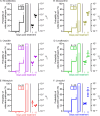
Outgrowth of intracellular S. aureus from a non-culturable state. SaOS-2 osteocyte-like cells were infected with S. aureus, and intracellular bacteria were confirmed non-culturable by 14 days post-infection. All cells were then treated for 3 days with either antibiotic-free media or with antibiotics at their respective 10× MIC. For all treatment groups (A: no antibiotic; B: doxycycline; C: oxacillin; D: levofloxacin; E: rifampicin; F: linezolid), cell culture media were tested for extracellular bacteria culturability from potential outgrowth on days 4, 7, and 11 (n = 32 wells per condition, line graphs on left Y-axis). Cell lysates from the experiment endpoint (day 11) were tested for intracellular bacterial culturability (n = 28 wells per condition, bar graphs on left Y-axis, hatched pattern indicates growth). The number of wells with or without outgrowth is shown adjacent. Total DNA isolated from cell lysates on days 1 and 11 was measured for intracellular bacterial genome levels by qPCR (n = 4 wells per condition; dot graphs on right Y-axis).

References

    1. Kavanagh N, Ryan EJ, Widaa A, Sexton G, Fennell J, O’Rourke S, Cahill KC, Kearney CJ, O’Brien FJ, Kerrigan SW. 2018. Staphylococcal osteomyelitis: disease progression, treatment challenges, and future directions. Clin Microbiol Rev 31:1–25. doi:10.1128/CMR.00084-17 - DOI - PMC - PubMed
    1. Davis JS, Metcalf S, Clark B, Robinson JO, Huggan P, Luey C, McBride S, Aboltins C, Nelson R, Campbell D, et al. . 2022. Predictors of treatment success after periprosthetic joint infection: 24-month follow up from a multicenter prospective observational cohort study of 653 patients. Open Forum Infect Dis 9:fac048. doi:10.1093/ofid/ofac048 - DOI - PMC - PubMed
    1. Lew DP, Waldvogel FA. 2004. Osteomyelitis. Lancet 364:369–379. doi:10.1016/S0140-6736(04)16727-5 - DOI - PubMed
    1. Tande AJ, Osmon DR, Greenwood-Quaintance KE, Mabry TM, Hanssen AD, Patel R. 2014. Clinical characteristics and outcomes of prosthetic joint infection caused by small colony variant staphylococci. MBio 5:e01910–14. doi:10.1128/mBio.01910-14 - DOI - PMC - PubMed
    1. Fraunholz M, Sinha B. 2012. Intracellular Staphylococcus aureus: live-in and let die. Front Cell Infect Microbiol 2:43. doi:10.3389/fcimb.2012.00043 - DOI - PMC - PubMed

Publication types

MeSH terms

LinkOut - more resources