. 2024 Jul 24;13(8):560.
doi: 10.3390/biology13080560.

Plasticity Comparison of Two Stem Cell Sources with Different Hox Gene Expression Profiles in Response to Cobalt Chloride Treatment during Chondrogenic Differentiation

Affiliations

Plasticity Comparison of Two Stem Cell Sources with Different Hox Gene Expression Profiles in Response to Cobalt Chloride Treatment during Chondrogenic Differentiation

Sahar Khajeh et al. Biology (Basel). .

Abstract

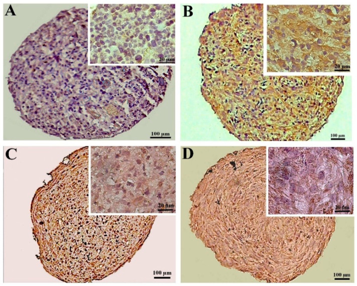
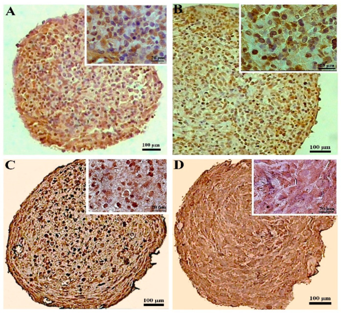
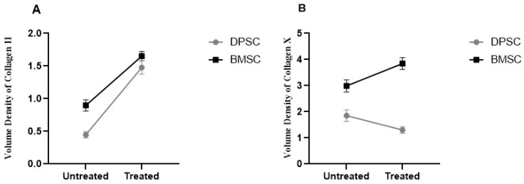
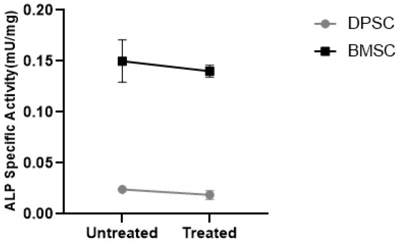
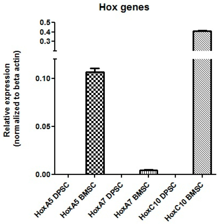
The limited self-repair capacity of articular cartilage is a challenge for healing injuries. While mesenchymal stem/stromal cells (MSCs) are a promising approach for tissue regeneration, the criteria for selecting a suitable cell source remain undefined. To propose a molecular criterion, dental pulp stem cells (DPSCs) with a Hox-negative expression pattern and bone marrow mesenchymal stromal cells (BMSCs), which actively express Hox genes, were differentiated towards chondrocytes in 3D pellets, employing a two-step protocol. The MSCs' response to preconditioning by cobalt chloride (CoCl2), a hypoxia-mimicking agent, was explored in an assessment of the chondrogenic differentiation's efficiency using morphological, histochemical, immunohistochemical, and biochemical experiments. The preconditioned DPSC pellets exhibited significantly elevated levels of collagen II and glycosaminoglycans (GAGs) and reduced levels of the hypertrophic marker collagen X. No significant effect on GAGs production was observed in the preconditioned BMSC pellets, but collagen II and collagen X levels were elevated. While preconditioning did not modify the ALP specific activity in either cell type, it was notably lower in the DPSCs differentiated pellets compared to their BMSCs counterparts. These results could be interpreted as demonstrating the higher plasticity of DPSCs compared to BMSCs, suggesting the contribution of their unique molecular characteristics, including their negative Hox expression pattern, to promote a chondrogenic differentiation potential. Consequently, DPSCs could be considered compelling candidates for future cartilage cell therapy.

Keywords: Hox genes; bone marrow mesenchymal stromal cell; chondrogenic differentiation; cobalt chloride; dental pulp stem cell; stem cell plasticity.


Conflict of interest statement

The authors declare no conflicts of interest.

Figures

Figure 1
Figure 1
Fibroblast-like morphology of passages 4–6 of the DPSCs (A) and BMSCs (B) in a monolayer culture, forming swirls at high confluency. Each arrow shows a representative stem cell.
Figure 2
Figure 2
Cell viability assay for the different CoCl2 concentrations (A) and exposure times (B). All treatments were compared to the control groups and represented as percentages of viability. ***, p < 0.001.
Figure 3
Figure 3
Gross morphology of the chondrogenic pellets at the initiation of differentiation (A), the round morphology at the end of differentiation (B), and the approximate size (C).
Figure 4
Figure 4
Tissue structures of the pellets stained by hematoxylin and eosin at day 21 of differentiation in the untreated (A,C) and CoCl2-pretreated (B,D) pellets derived from the DPSCs (A,B) and BMSCs (C,D). The lacuna-like structures are shown with black arrows.
Figure 5
Figure 5
GAGs staining by alcian blue at day 21 of differentiation in the untreated (A,C) and CoCl2-pretreated (B,D) samples for the DPSCs (A,B) and BMSCs chondrogenic pellets (C,D). A lacuna-like structure is shown with a black arrow.
Figure 6
Figure 6
Immunostaining for collagen II at day 21 of chondrogenic induction in the untreated (A,C) and CoCl2-pretreated (B,D) pellets derived from the DPSCs (A,B) and BMSCs (C,D).
Figure 7
Figure 7
Immunostaining for collagen X at day 21 of chondrogenic induction in the untreated (A,C) and CoCl2-pretreated (B,D) pellets derived from the DPSCs (A,B) and BMSCs (C,D).
Figure 8
Figure 8
Volume densities of collagen II (A) and collagen X (B) in the untreated and CoCl2-pretreated pellets.
Figure 9
Figure 9
ICP-MS for the assessment of CoCl2 uptake by the DPSCs and BMSCs after 100 μM of CoCl2 treatment for 24 h. **, p < 0.01.
Figure 10
Figure 10
The GAGs contents were shown as the absorbance of the eluted alcian blue compared to the DNA content (ng) ratio.
Figure 11
Figure 11
ALP specific activity in the MSC chondrogenic pellets represented as mU/mg.
Figure 12
Figure 12
mRNA expression of the Hox genes in the DPSCs and BMSCs.

Similar articles

References

    1. Samvelyan H.J., Hughes D., Stevens C., Staines K.A. Models of osteoarthritis: Relevance and new insights. Calcif. Tissue Int. 2021;109:243–256. doi: 10.1007/s00223-020-00670-x. - DOI - PMC - PubMed
    1. Khajeh S., Bozorg-Ghalati F., Zare M., Panahi G., Razban V. Cartilage tissue and therapeutic strategies for cartilage repair. Curr. Mol. Med. 2021;21:56–72. doi: 10.2174/1566524020666200610170646. - DOI - PubMed
    1. Malekpour K., Hazrati A., Zahar M., Markov A., Zekiy A.O., Navashenaq J.G., Roshangar L., Ahmadi M. The potential use of mesenchymal stem cells and their derived exosomes for orthopedic diseases treatment. Stem Cell Rev. Rep. 2022;18:933–951. doi: 10.1007/s12015-021-10185-z. - DOI - PMC - PubMed
    1. Asadi-Golshan R., Razban V., Mirzaei E., Rahmanian A., Khajeh S., Mostafavi-Pour Z., Dehghani F. Efficacy of dental pulp-derived stem cells conditioned medium loaded in collagen hydrogel in spinal cord injury in rats: Stereological evidence. J. Chem. Neuroanat. 2021;116:101978. doi: 10.1016/j.jchemneu.2021.101978. - DOI - PubMed
    1. Hodge J.G., Decker H.E., Robinson J.L., Mellott A.J. Tissue-Mimetic Culture Enhances Mesenchymal Stem Cell Secretome Capacity to Improve Regenerative Activity of Keratinocytes and Fibroblasts in vitro. Wound Repair Regen. 2023;31:367–383. doi: 10.1111/wrr.13076. - DOI - PubMed

LinkOut - more resources