. 2024 Aug 8;14(8):383.
doi: 10.3390/bios14080383.

Microbead-Encapsulated Luminescent Bioreporter Screening of P. aeruginosa via Its Secreted Quorum-Sensing Molecules

Affiliations

Microbead-Encapsulated Luminescent Bioreporter Screening of P. aeruginosa via Its Secreted Quorum-Sensing Molecules

Abraham Abbey Paul et al. Biosensors (Basel). .

Abstract

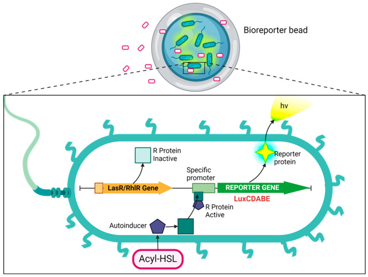
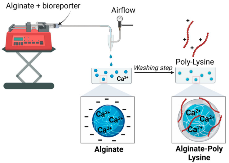
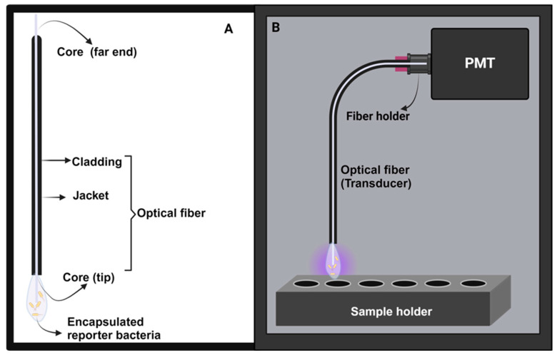
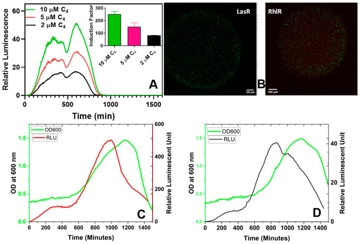
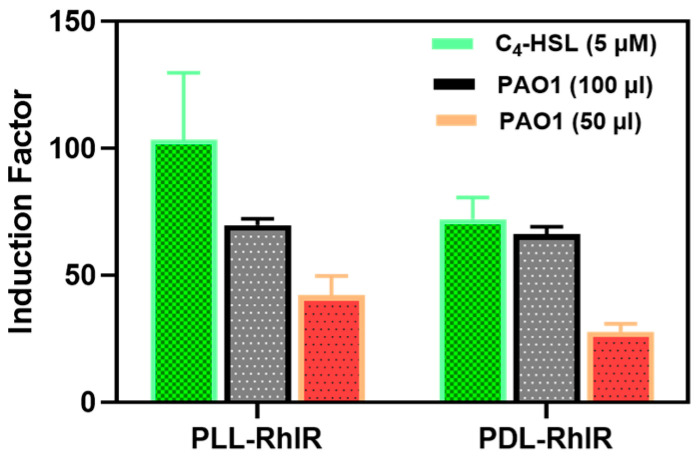
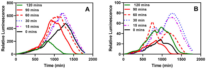
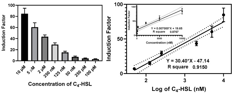
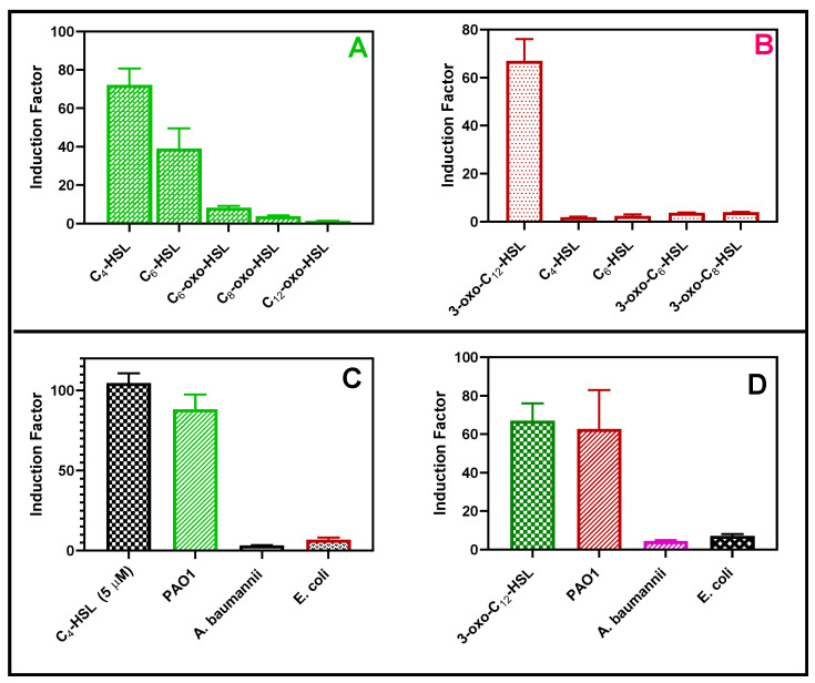
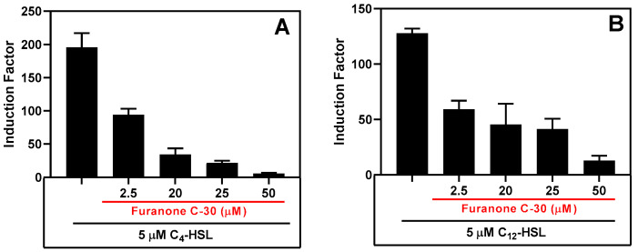
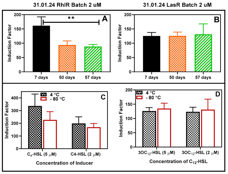
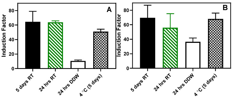
Pseudomonas aeruginosa is an opportunistic Gram-negative bacterium that remains a prevalent clinical and environmental challenge. Quorum-sensing (QS) molecules are effective biomarkers in pinpointing the presence of P. aeruginosa. This study aimed to develop a convenient-to-use, whole-cell biosensor using P. aeruginosa reporters individually encapsulated within alginate-poly-L-lysine (alginate-PLL) microbeads to specifically detect the presence of bacterial autoinducers. The PLL-reinforced microbeads were prepared using a two-step method involving ionic cross-linking and subsequent coating with thin layers of PLL. The alginate-PLL beads showed good stability in the presence of a known cation scavenger (sodium citrate), which typically limits the widespread applications of calcium alginate. In media containing synthetic autoinducers-such as N-(3-oxo dodecanoyl) homoserine lactone (3-oxo-C12-HSL) and N-butanoyl-L-homoserine lactone (C4-HSL), or the cell-free supernatants of planktonic or the flow-cell biofilm effluent of wild P. aeruginosa (PAO1)-the encapsulated bacteria enabled a dose-dependent detection of the presence of these QS molecules. The prepared bioreporter beads remained stable during prolonged storage at 4 and -80 °C and were ready for on-the-spot sensing without the need for recovery. The proof-of-concept, optical fiber-based, and whole-cell biosensor developed here demonstrates the practicality of the encapsulated bioreporter for bacterial detection based on specific QS molecules.

Keywords: 3-oxo-C12-HSL; C4-HSL; Furanone C-30; Pseudomonas aeruginosa; QS inhibitor; alginate; autoinducers; bioencapsulation; hydrogels; microbeads; optical biosensors; poly-lysine; quorum sensing; whole-cell biosensors.


Conflict of interest statement

R.S.M. is the co-founder of footprints. The other authors declare no conflicts of interest.

Figures

Figure 1
Figure 1
Schematic of the cellular events that result in the expression of a reporter protein. The cell can easily take up a bioavailable analyte, such as AHL, through its membrane. AHL (3-oxo-C12-HSL and C4-HSL) binds with a regulatory protein called LasR or RhlR, forming a protein–AHL complex. This complex binds to its corresponding promoter, Plasl or Prhl, which triggers activation of the transcription and translation of a reporter gene called luxCDABE. This results in the expression of the reporter protein, luciferase, inducing the bioluminescent reaction characterized by the emission of blue-green light (~490 nm). Created with BioRender.com, accessed on 13 June 2024.
Figure 2
Figure 2
Schematics of the microcapsule synthesis. The size of the beads can be controlled by changing the flow rate of the feed solution (alginate + bioreporter) and the air pressure.
Figure 3
Figure 3
The basic components of the whole-cell, fiber-optic biosensors. (A) Optical fiber with a bioreporter immobilized unto the core. (B) Light–tight portable black box encasing all the biosensor components. For the image of the black box, readers are directed to the Supplementary Materials section (Figure S19).
Figure 4
Figure 4
The ATR-FTIR absorbance spectra of the outer dehydrated alginate-PPL microcapsules. (a) The 1700–900 cm−1 region of the spectra that corresponds to the asymmetric and symmetric stretching vibrations characteristics of the carboxyl (COO−1) functional group. (b) The full spectra of ca-alginate, alginate-PLL, and the characteristic peaks are discussed in the text. A spectrum of the alginate/PLL interpenetrating network and sodium alginate can be found in the Supplementary Materials section (Figure S5).
Figure 5
Figure 5
The deconvoluted XPS spectrum peaks of the lyophilized alginate-poly-lysine beads. The deconvoluted peaks were assigned to a chemical group based on the binding energy of the peaks (N1s, O1s, C1s, and Ca2p), as shown in Figure S6.
Figure 6
Figure 6
Scanning electron microscopy (SEM) images of the outer part of the alginate and alginate-poly-lysine microbeads at different magnifications. (A) Alginate microbeads prior to the poly-lysine (PLL) coating. (B) Alginate-PLL microbeads. (C) The alginate/PLL interpenetrating network (IPN) prior to the PLL outer coating. (D) PLL-coated alginate/PLL IPN.
Figure 7
Figure 7
Stability of alginate-poly-lysine microbeads in the presence of cation scavengers. (A) The alginate microbeads (control) were rapidly degraded within 60 min when incubated in 5% w/v sodium citrate because of the Ca2+ removal from the hydrogels. (B) Alginate-PLL. (C) The alginate-PLL IPN capsules remained stable in a 5% sodium citrate solution after 14 h.
Figure 8
Figure 8
Swelling properties. (A) beads stored in refrigerators (pristine), air-dried, and rehydrated (wet). (B) beads stored at −80 °C (pristine), air-dried and rehydrated (wet). (C) The physical appearance of the beads during wet, dried, and swelled conditions.
Figure 9
Figure 9
The bioluminescent response of the immobilized RhlR in different C4-HSL concentrations. The insert in (A) illustrates the induction factor calculated using the maximum relative luminescence unit (RLU) derived from the spectral data. The induction factor is the ratio of the test’s maximum RLU conducted in the presence of an inducer to the maximum RLU obtained in the absence of added inducers. (B) Confocal images of the live/dead stained bacteria within alginate-PLL beads. (C) Both the luminescence and cell density (absorbance at 600 nm) of the LasR bioreporter. (D) The luminescence and OD600 readings of the RhlR bioreporter. The OD600-normalized luminescence reading (RLU/OD600) can be found in Figure S8.
Figure 10
Figure 10
The bioluminescent responses of the PLL- and PDL-coated alginate were similar. The PDL and PLL coatings were compared regarding the bacteria’s response to the added synthetic (C4-HSL) and secreted (by the PAO1 wild strain of P. aeruginosa) autoinducers.
Figure 11
Figure 11
The effect of pre-incubation and shaking before testing. (A) Bioluminescent response of the LasR strain. (B) Bioluminescent response of the RhlR strain. The stored microspheres were analyzed with or without pre-incubation to determine the effect of pre-incubation on the biosensor performance of the reporter bacteria strains. Experiments were conducted with 0, 15, 30, 60, 90, and 120 min of incubation and shaking at 37 °C and 220 rpm. Each curve represents the mean of four replicate experiments.
Figure 12
Figure 12
Calibration curve of the RlhR bioreporter beads at various concentrations of C4-HSL. The left panel shows the bioluminescence (expressed as an induction factor) produced by the encapsulated reporter in the presence of increasing concentrations of exogenously added C4-HSL. The right panel shows the dose–response relationships inferred from the data in the left panel. Linear regression lines (bold) were drawn on logarithmic and linear (upper insert) scales, and the 95% confidence interval limits were the black dotted lines parallel to the regression lines. The equation and the R2 value for each regression line are shown. Data are the mean ± SD of three independent experiments.
Figure 13
Figure 13
The bioreporter specificity test. The RhlR (A) and LasR (B) response toward 5 µM each of synthetic QS molecules, respectively. The second experiment was conducted with either 20 µL of cell-free supernatants of each Gram-negative bacterium such as Escherichia coli (E. coli), Pseudomonas aeruginosa (PAO1), and Acinetobacter baumannii (A. baumannii) (72 h biofilm set up), or 5 µM of C4-HSL for the RhlR strain (C) or 5 µM of 3-oxo-C12-HSL for the LasR strain (D).
Figure 14
Figure 14
The QS inhibitory effect of furanone C-30 in the presence of synthetic AHLs. (A) The inhibition of RhlR signaling in the presence of 5 µM of C4-HSL and (B) the inhibition of the LasR bioluminescence system in the presence of 5 µM of 3-OC12-HSL. Luminescent readings were taken for 10 h, and the maximum relative light unit (RLU) was plotted. The results are the mean ± SD of biological triplicates.
Figure 15
Figure 15
Storability of the bioreporter beads in refrigerator conditions. (A) RhlR and (B) LasR 2 µM inducers. The RhlR strain experienced a significant (shown as ** at p < 0.05) reduction in activities between Days 7 and 50 and remained fairly constant thereafter. Comparison of the effect of storage at 4 and −80 degrees (for 30 days) on the luminescence. The residual activity of RhlR (C) and LasR (D) was compared after storage in the refrigerator and freezer at −80 °C for a minimum of 30 days. Two concentrations of each AHL were tested, and the results are reported as the mean ± SD, n = 4. Statistical analysis revealed no significant differences in the readings at p < 0.05.
Figure 16
Figure 16
Storage at room temperature. (A) RhlR and (B) LasR results. The results are presented as the mean ± SD, (n = 4). RT: beads stored dried at room temperature.

Similar articles

Cited by

References

    1. Van Delden C., Iglewski B.H. Cell-to-cell signaling and Pseudomonas aeruginosa infections. Emerg. Infect. Dis. 1998;4:551. doi: 10.3201/eid0404.980405. - DOI - PMC - PubMed
    1. Lyczak J.B., Cannon C.L., Pier G.B. Lung infections associated with cystic fibrosis. Clin. Microbiol. Rev. 2002;15:194–222. doi: 10.1128/CMR.15.2.194-222.2002. - DOI - PMC - PubMed
    1. Kazemian H., Ghafourian S., Heidari H., Amiri P., Yamchi J.K., Shavalipour A., Houri H., Maleki A., Sadeghifard N. Antibacterial, anti-swarming and anti-biofilm formation activities of Chamaemelum nobile against Pseudomonas aeruginosa. Rev. Soc. Bras. Med. Trop. 2015;48:432–436. doi: 10.1590/0037-8682-0065-2015. - DOI - PubMed
    1. Winson M.K., Camara M., Latifi A., Foglino M., Chhabra S.R., Daykin M., Bally M., Chapon V., Salmond G., Bycroft B.W. Multiple N-acyl-L-homoserine lactone signal molecules regulate production of virulence determinants and secondary metabolites in Pseudomonas aeruginosa. Proc. Natl. Acad. Sci. USA. 1995;92:9427–9431. doi: 10.1073/pnas.92.20.9427. - DOI - PMC - PubMed
    1. Middleton B., Rodgers H.C., Cámara M., Knox A.J., Williams P., Hardman A. Direct detection of N-acylhomoserine lactones in cystic fibrosis sputum. FEMS Microbiol. Lett. 2002;207:1–7. doi: 10.1111/j.1574-6968.2002.tb11019.x. - DOI - PubMed

LinkOut - more resources