. 2024 Jul 28;15(8):574.
doi: 10.3390/insects15080574.

Sensillar Ultrastructure of the Antennae and Maxillary Palps of the Warble Fly Oestromyia leporina (Pallas, 1778) (Diptera: Oestridae)

Affiliations

Sensillar Ultrastructure of the Antennae and Maxillary Palps of the Warble Fly Oestromyia leporina (Pallas, 1778) (Diptera: Oestridae)

Zhuowei An et al. Insects. .

Abstract

Despite the development of molecular techniques, morphological phylogeny still remains integral in underpinning the relationship between some clades of Calyptratae, especially the ones with fast radiation, such as those in Oestridae (Diptera: Brachycera), yet few synapomorphy has been proposed for adults in this family. Using scanning electron microscopy, we investigated the morphological structure and ultrastructure of the antennae and maxillary palps of adult Oestromyia leporina (Hypodermatinae, Oestridae). One type of trichoid sensillum (Tr), three types of basiconic sensilla (Ba I, Ba II, and Ba III), one type of coeloconic sensillum (Co I), and one type of clavate sensillum (Cl) were found on the antennal postpedicel. Surprisingly, this species has the most complex types of sensilla on the maxillary palps that have been reported in Calyptratae so far, with two types of coeloconic sensilla (Co II and Co III) and two types of mechanoreceptors. We then identified three common characteristics on the arista of Oestridae (Hypodermatinae, Oestrinae, Gasterophilinae and Cuterebrinae) that are potential synapomorphies. These characteristics indicate the value of the morphology of maxillary palps and aristae in taxonomy studies of Calyptratae.

Keywords: Oestromyia leporina; antenna; maxillary palps; morphology; sensilla; sensory pits; ultrastructure.


Conflict of interest statement

The authors declare no conflicts of interest.

Figures

Figure 1
Figure 1
Scanning electron micrographs of general morphology of antenna in Oestromyia leporina. (A) Dorsolateral and posteroventral surface of antenna. (B) Anterodorsal surface of antenna. (C) Anterodorsal surface of antennal scape and pedicel, showing the distribution of mechanoreceptors; box shows the longitudinal grooves on mechanoreceptors. (D) Morphology of antennal pedicel after removal of the postpedicel. Scale bars: (A) 160 μm, (B) 200 μm, (C) 100 μm, 50 μm in inset, (D) 50 μm.
Figure 2
Figure 2
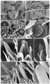
Scanning electron micrographs of general morphology and the sensilla of maxillary palps in Oestromyia leporina. (A) General view of maxillary palps and beak. (B) General view of the right maxillary palp. (C) Ba I and Cl in sensory pits on maxillary palp surface. (D) Co III, arrow indicates the pore on the sensillar surface. (E) General view of Ba I. (F,G) General view of Cl. (HJ) Co II, showing it is much longer than Co I; arrow indicates only one pore on the top of the sensilla. Scale bars: (A) 150 μm, (B) 100 μm, (C) 10 μm, (D) 3 μm, (E) 5 μm, (F) 5 μm, (G) 2.5 μm, (H) 5 μm, (I) 5 μm, (J) 2 μm. Abbreviations: Me I, mechanoreceptors I; Me II, mechanoreceptors II; Ba I, basiconic sensilla I; Co I, coeloconic sensilla I; Co II, coeloconic sensilla II; Co III, coeloconic sensilla III; Cl, clavate sensilla.
Figure 3
Figure 3
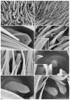
Scanning electron micrographs of general morphology of the sensilla on postpedicel in Oestromyia leporina. (A,B) Distribution of sensilla on the postpedicel surface. (C) Ba II, arrow shows two large pores on the end. (D) Tr, box shows pores on its surface. (E) Ba I, pores can be observed on the cuticular wall. (F) Co I, set in shallow depression on postpedicel surface. (G) Cl with numerous pores on the cuticular wall. (H) Cl with protrusions on the top. Scale bars: (A) 10 μm, (B) 10 μm, (C) 1 μm, (D) 5 μm, 0.5 μm in inset, (E) 2.5 μm, (F) 1.5 μm, 1 μm in inset, (G) 1 μm, (H) 2.5 μm. Abbreviations: Tr, trichoid sensilla; Ba I, basiconic sensilla I; Ba II, basiconic sensilla II; Co I, coeloconic sensilla I; Cl, clavate sensilla.
Figure 4
Figure 4
Scanning electron micrographs of the arista in Oestromyia leporina. (A,B) The proximal end of the arista, showing segmented and enlarged base. (C) The distal end of the arista, showing the twisted tip. (D) The base of the arista, arrows indicate Co I. (E) Ba III on the base of the arista, arrows indicate Ba III. (F) Enlarged view of Ba III. Scale bars: (A) 50 μm, (B) 20 μm, (C) 50 μm, (D) 15 μm, (E) 10 μm, (F) 1 μm. Abbreviations: Ba III, basiconic sensilla III; Co I, coeloconic sensilla I.
Figure 5
Figure 5
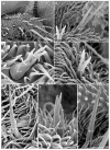
Scanning electron micrographs of sensory pits in Oestromyia leporina. (A,B) Sensory pits on postpedicel surface. (C,D) Enlarged view of a sensory pit, housing multiple Cl. (EG) Lateral view of Ba I, Cl, and Co I in sensory pits. Scale bars: (A) 10 μm, (B) 30 μm, (C) 10 μm, (D) 10 μm, (E) 10 μm, (F) 2.5 μm, (G) 5 μm. Abbreviations: Ba I, basiconic sensilla I; Co I, coeloconic sensilla I; Cl, clavate sensilla.
Figure 6
Figure 6
Scanning electron micrographs of mechanoreceptors on the maxillary palps in Oestromyia leporina. (A) Overview of the mechanoreceptors on maxillary palps. (B,D) Bifurcated mechanoreceptors with ridges on surface. (C) A short trifurcated mechanoreceptor with smooth surface. (E) A bifurcated mechanoreceptor with two long branches. (F) A trifurcated mechanoreceptor with smooth surface. (G) A long trifurcated mechanoreceptor with ridges on the surface. Scale bars: (A) 25 μm, (B) 10 μm, (C) 5 μm, (D) 10 μm, (E) 10 μm, (F) 10 μm, (G) 15 μm. Abbreviations: Me I, mechanoreceptors I; Me II, mechanoreceptors II.

References

    1. Kutty S.N., Pape T., Wiegmann B.M., Meier R. Molecular phylogeny of the Calyptratae (Diptera: Cyclorrhapha) with an emphasis on the superfamily Oestroidea and the position of Mystacinobiidae and McAlpine’s fly. Syst. Entomol. 2010;35:614–635. doi: 10.1111/j.1365-3113.2010.00536.x. - DOI
    1. Li X., Cai X.D., Ding S.M., Wang L., Li W.L., Liu X.Y., Zhang C.T., Yang D. Phylogeny and evolutionary timescale of muscidae (Diptera: Calyptratae) inferred from mitochondrial genomes. Insects. 2023;14:286. doi: 10.3390/insects14030286. - DOI - PMC - PubMed
    1. Yan L.P., Xu W.T., Zhang D., Li J.Q. Comparative analysis of the mitochondrial genomes of flesh flies and their evolutionary implication. Int. J. Biol. Macromol. 2021;174:385–391. doi: 10.1016/j.ijbiomac.2021.01.188. - DOI - PubMed
    1. Yan L.P., Pape T., Elgar M., Gao Y.Y., Zhang D. Evolutionary history of stomach bot flies in the light of mitogenomics. Syst. Entomol. 2019;44:797–809. doi: 10.1111/syen.12356. - DOI
    1. Lee M.S., Palci A. Morphological phylogenetics in the genomic age. Curr. Biol. CB. 2015;25:922–929. doi: 10.1016/j.cub.2015.07.009. - DOI - PubMed

Grants and funding

LinkOut - more resources