. 2024 Aug 12;10(8):567.
doi: 10.3390/jof10080567.

A Chimeric ORF Fusion Phenotypic Reporter for Cryptococcus neoformans

Affiliations

A Chimeric ORF Fusion Phenotypic Reporter for Cryptococcus neoformans

Louis S Phillips-Rose et al. J Fungi (Basel). .

Abstract

The plethora of genome sequences produced in the postgenomic age has not resolved many of our most pressing biological questions. Correlating gene expression with an interrogatable and easily observable characteristic such as the surrogate phenotype conferred by a reporter gene is a valuable approach to gaining insight into gene function. Many reporters including lacZ, amdS, and the fluorescent proteins mRuby3 and mNeonGreen have been used across all manners of organisms. Described here is an investigation into the creation of a robust, synthetic, fusion reporter system for Cryptococcus neoformans that combines some of the most useful fluorophores available in this system with the versatility of the counter-selectable nature of amdS. The reporters generated include multiple composition and orientation variants, all of which were investigated for differences in expression. Evaluation of known promoters from the TEF1 and GAL7 genes was undertaken, elucidating novel expression tendencies of these biologically relevant C. neoformans regulators of transcription. Smaller than lacZ but providing multiple useful surrogate phenotypes for interrogation, the fusion ORF serves as a superior whole-cell assay compared to traditional systems. Ultimately, the work described here bolsters the array of relevant genetic tools that may be employed in furthering manipulation and understanding of the WHO fungal priority group pathogen C. neoformans.

Keywords: Cryptococcus neoformans; amdS; fluorescent protein; fungal pathogen; fusion gene; reporter.


Conflict of interest statement

The authors declare no conflicts of interest.

Figures
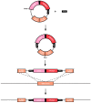
Figure 1
Figure 1
Schematic overview of the bicistronic reporter construct integrating into C. neoformans Safe Haven locus. Built into the pSDMA57 plasmid backbone, engineered to be linearised using PacI endonuclease. The amdS-mRuby3 bicistronic reporter construct utilises the 5′ and 3′ flanking regions of the Safe Haven as regions of homologous recombination for integration. Expression is driven by the constitutive promoter of TEF1, which was inserted using the SwaI endonuclease site. The neomycin resistance gene (NEO) present in the plasmid backbone was used as the selectable marker to identify successful gene deletion mutants.
Figure 2
Figure 2
Only the first of two ORFs is expressed in the bicistronic reporter construct. Spotting assay of amdS:mRuby3 bicistronic strain on 2% glucose YNB agar media plates supplemented as depicted. Strains individually expressing amdS or mRuby3 were used as controls. mRuby3 was excited using 535 nm light, capturing fluorescence between 565–645 nm. The wild-type control is H99O. Plates were photographed following 72 h of incubation at 30 °C.
Figure 3
Figure 3
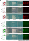
Fusing two ORFs produces reporters with multiple useful phenotypes. (A) Spotting assay of amdS:mRuby3 fusion strain on 2% glucose YNB agar media plates supplemented as depicted. mRuby3 was excited using 535 nm light, capturing fluorescence between 565 and 645 nm. (B) Spotting assay of amdS:mNeonGreen fusion strain on 2% glucose YNB agar media plates supplemented as depicted. mNeonGreen was excited using 460 nm light, capturing fluorescence between 505 and 545 nm. Strains individually expressing amdS, mRuby3 or mNeonGreen were used as controls. The wild-type control is H99O. Plates were photographed following 72 h of incubation at 30 °C.
Figure 4
Figure 4
Order of ORF fusion does not affect phenotypic output for the fusion reporter when constitutively expressed using TEF1(p). (A) Spotting assay of mRuby3:amdS fusion strain on 2% glucose YNB agar media plates supplemented as depicted. mRuby3 was excited using 535 nm light, capturing fluorescence between 565 and 645 nm. (B) Spotting assay of mNeonGreen:amdS fusion strain on 2% glucose YNB agar media plates supplemented as depicted. mNeonGreen was excited using 460 nm light, capturing fluorescence between 505 and 545 nm. Strains individually expressing amdS, mRuby3 or mNeonGreen were used as controls. The wild-type control is H99O. Plates were photographed following 72 h of incubation at 30 °C.
Figure 5
Figure 5
Order of ORF fusion affects fluorescence phenotype for the fusion reporter when expressed using inducible GAL7(p). Both orientations of the mRuby3 and mNeonGreen dual fusion reporters driven by GAL7 promoter plated on supplemented YNB agar. Controls are wild-type H99O and the four reporter variants under the control of the constitutive TEF1 promoter. Plates were photographed following 72 h of incubation at 30 °C. C. neoformans reporter strains in a spotting assay on 2% glucose, 2% glucose + 2% galactose, or 2% galactose. Strains were grown on media containing the indicated supplements. (A) mRuby3 was excited using 535 nm light, capturing fluorescence between 565 and 645 nm. (B) mNeonGreen was excited using 460 nm light, capturing fluorescence between 505 and 545 nm.
Figure 6
Figure 6
Fluorescence of fusion reporters evaluates inducible promoter expression. GAL7(p)-fluorophore:amdS fusion reporter strains were exposed to galactose-supplemented liquid YNB media following growth on 2% glucose. C. neoformans cells were incubated over 12 h and sampled at hourly time points for flow cytometry analysis. Relative fluorescence was calculated using maximal TEF1(p)-fluorophore:amdS strains run through CytoFlexS flow cytometry machine. Cell count (histogram) was standardised (20,000 cells) and relative fluorescence was recorded and displayed for each time point. Mean fluorescent intensity (MFI) was calculated using FlowJo analysis software. Data were collected across five biological replicates. Gating methodology is detailed in Supplementary Figure S2. (A) mRuby3 fluorescence was captured with a 610 ± 10 nm bandpass filter. (B) mNeonGreen fluorescence was captured with a 510 ± 20 nm bandpass filter.

Similar articles

References

    1. Fleischmann R.D., Adams M.D., White O., Clayton R.A., Kirkness E.F., Kerlavage A.R., Bult C.J., Tomb J.-F., Dougherty B.A., Merrick J.M., et al. Whole-Genome Random Sequencing and Assembly of Haemophilus influenzae Rd. Science. 1995;269:496–512. doi: 10.1126/science.7542800. - DOI - PubMed
    1. Goffeau A., Barrell B.G., Bussey H., Davis R.W., Dujon B., Feldmann H., Galibert F., Hoheisel J.D., Jacq C., Johnston M., et al. Life with 6000 Genes. Science. 1996;274:546–567. doi: 10.1126/science.274.5287.546. - DOI - PubMed
    1. Sanger F., Air G.M., Barrell B.G., Brown N.L., Coulson A.R., Fiddes C.A., Hutchison C.A., Slocombe P.M., Smith M. Nucleotide Sequence of Bacteriophage Phi X174 DNA. Nature. 1977;265:687–695. doi: 10.1038/265687a0. - DOI - PubMed
    1. Venter J.C., Adams M.D., Myers E.W., Li P.W., Mural R.J., Sutton G.G., Smith H.O., Yandell M., Evans C.A., Holt R.A., et al. The Sequence of the Human Genome. Science. 2001;291:1304–1351. doi: 10.1126/science.1058040. - DOI - PubMed
    1. Lander E.S., Linton L.M., Birren B., Nusbaum C., Zody M.C., Baldwin J., Devon K., Dewar K., Doyle M., FitzHugh W., et al. Initial Sequencing and Analysis of the Human Genome. Nature. 2001;409:860–921. doi: 10.1038/35057062. - DOI - PubMed

LinkOut - more resources