Review
. 2024 Jul 29;10(8):182.
doi: 10.3390/jimaging10080182.

Special Types of Breast Cancer: Clinical Behavior and Radiological Appearance

Affiliations
Review

Special Types of Breast Cancer: Clinical Behavior and Radiological Appearance

Marco Conti et al. J Imaging. .

Abstract

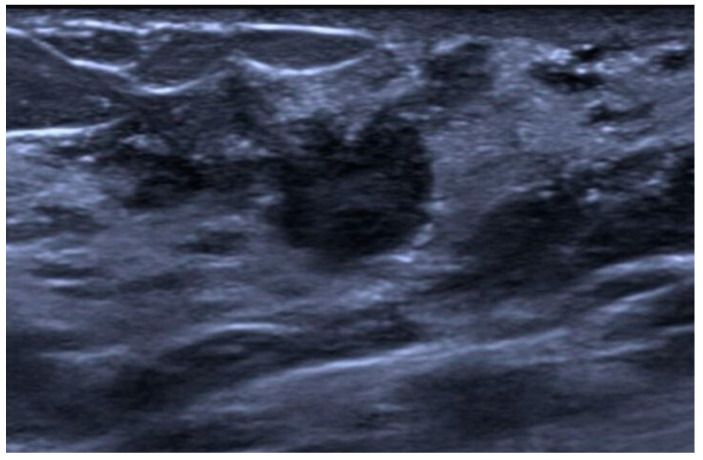
Breast cancer is a complex disease that includes entities with different characteristics, behaviors, and responses to treatment. Breast cancers are categorized into subgroups based on histological type and grade, and these subgroups affect clinical presentation and oncological outcomes. The subgroup of "special types" encompasses all those breast cancers with insufficient features to belong to the subgroup "invasive ductal carcinoma not otherwise specified". These cancers account for around 25% of all cases, some of them having a relatively good prognosis despite high histological grade. The purpose of this paper is to review and illustrate the radiological appearance of each special type, highlighting insights and pitfalls to guide breast radiologists in their routine work.

Keywords: breast cancer; breast imaging; conventional and advanced imaging; invasive lobular carcinoma; special types.


Conflict of interest statement

The authors declare no conflicts of interest.

Figures

Figure 1
Figure 1
Invasive lobular carcinoma in a 53-year-old patient. (a) Cranio-caudal and (b) medio-lateral oblique mammograms of the left breast show an architectural distortion in the upper-outer quadrant, (c) corresponding to an irregular-shaped hypoechoic lesion with spiculated margins on ultrasound. (d) Axial and (e) sagittal T1-weighted fat-suppressed dynamic contrast-enhanced MRI images reveal an irregular mass with spiculated margins in the upper-outer quadrant.
Figure 2
Figure 2
Invasive lobular carcinoma in a 78-year-old patient. Contrast-enhancement mammography: (a) low-energy cranio-caudal and (c) medio-lateral oblique images of the left breast show a high-density irregular mass, (b,d) corresponding to an area of mass enhancement on the recombined images (white arrow). Recombined images highlight the presence of three satellite nodules (white arrowheads), demonstrating the multicentricity of the disease. (e) US image shows an irregular hypoechoic mass with spiculated margins corresponding to the primary lesion, indicated by the white arrow in (b,d).
Figure 3
Figure 3
Tubular carcinoma. (a) Cranio-caudal and (b) medio-lateral oblique 2D synthetic mammograms (c), cranio-caudal and (d) medio-lateral oblique DBT images, and (e) spot compression view of the left breast showing a small opacity (f) corresponding to a hypoechoic mass with non-circumscribed margins on ultrasound.
Figure 4
Figure 4
Mucinous carcinoma in a 58-year-old patient presenting with a right-breast lump on clinical examination. (a) Cranio-caudal and (b) medio-lateral oblique mammograms of the right breast show an oval-shaped mass with circumscribed margins at the junction of the upper and lower outer quadrants (c) corresponding to a hypoechoic lesion on ultrasound. (d) Axial T2-weighted MRI image shows multiple areas of high signal intensity within the mass corresponding to the mucinous content. (e) Axial T1-weighted fat-suppressed dynamic contrast-enhanced MRI image demonstrates a heterogeneous internal enhancement.
Figure 5
Figure 5
Mucinous carcinoma in a 56-year-old patient. (a) Cranio-caudal and (b) medio-lateral oblique 2D synthetic mammograms; (c) cranio-caudal and (d) medio-lateral oblique DBT images of the left breast show a solid mass with circumscribed margins (e) corresponding on US to a mass with circumscribed margins and acoustic enhancement that could mimic a benign lesion (e.g., fibroadenoma).
Figure 6
Figure 6
Breast MRI of a 45-year-old patient with a mucinous carcinoma of the right breast. (a) Axial T2-weighted MRI image shows multiple mass-like lesions tightly bound one to each other, one in continuity with the skin, characterized by internal areas of high signal intensity. (b) Axial T1-weighted fat-suppressed contrast-enhanced dynamic image demonstrates a heterogeneous internal enhancement.
Figure 7
Figure 7
Mucinous cystadenocarcinoma in a 40-year-old patient. US image showing a complex cystic mass with a solid isoechoic component.
Figure 8
Figure 8
Medullary carcinoma in a 60-year-old patient. (a) Cranio-caudal and (b) medio-lateral oblique 2D synthetic mammograms, (c) cranio-caudal DBT image, (d) medio-lateral oblique DBT image and (e) spot compression view of the left breast showing an oval-shaped opacity in the upper-outer quadrant (f) corresponding to an oval-shaped homogeneous hypoechoic mass on ultrasound.
Figure 9
Figure 9
Papillary carcinoma in a 49-year-old patient. Contrast-enhancement mammography: low-energy (a) cranio-caudal and (c) medio-lateral oblique images of the right breast show a high-density mass, apparently oval-shaped (only partially included), at the junction of the upper and lower outer quadrants. Some microcalcifications are visible within the mass. (bd) The mass corresponds to an area of enhancement on the recombined images.
Figure 10
Figure 10
Papillary carcinoma in a 59-year-old patient. (a) Cranio-caudal and (b) medio-lateral oblique mammograms of the left breast show a round opacity in the lower-inner quadrant (c) corresponding on ultrasound to a complex cystic mass with a solid anterior component. A fluid−fluid level is visible inside the cyst due to the bleeding produced during the biopsy procedure. (d) Axial 3D gradient echo T1-weighted post-contrast MRI image of the left breast shows the enhancement of the solid component of the lesion.
Figure 11
Figure 11
Micropapillary carcinoma in a 75-year-old patient. US image shows a round hypoechoic mass with non-circumscribed margins.
Figure 12
Figure 12
Apocrine carcinoma in a 61-year-old patient. (a) Cranio-caudal and (b) medio-lateral oblique DBT images of the left breast show a small, irregularly shaped opacity in the lower-inner quadrant (c) corresponding to a hypoechoic mass with non-circumscribed margins on ultrasound.
Figure 13
Figure 13
Metaplastic carcinoma in a 43-year-old patient. (a) Cranio-caudal and (b) medio-lateral oblique mammograms of the right breast show a high-density mass with indistinct margins predominantly located in the upper-outer quadrant and (c) corresponding to an iso-hypoechoic mass with partially indistinct margins and cystic components on ultrasound.
Figure 14
Figure 14
Metaplastic carcinoma in a 40-year-old patient. (a) Medio-lateral oblique mammogram of the right breast shows a high-density mass, only partially included, with indistinct margins in the upper quadrant in the posterior third. Surgical clips from a previous surgery are visible. The cranio-caudal projection is not presented because the mass was not visible on it due to its position. (b) On ultrasound, it corresponds to a hypoechoic mass with irregular margins. (c) Axial T2-weighted MRI image shows high-signal-intensity areas within the mass corresponding to necrotic components, and (d) axial 3D gradient echo T1-weighted post-contrast MRI image demonstrates a ringlike uptake.
Figure 15
Figure 15
Solid-basaloid variant of adenoid cystic carcinoma. (a) Cranio-caudal and (b) medio-lateral oblique 2D synthetic mammograms, (c) cranio-caudal and (d) medio-lateral oblique DBT images of the right breast show a round opacity with circumscribed margins in the upper-outer quadrant (e,f) corresponding to a hypoechoic mass with cystic components on ultrasound. (g) Axial T2-weighted fat-suppressed MRI image shows high signal from the cystic components, and (h) axial T1-weighted fat-suppressed dynamic contrast-enhanced MRI image demonstrates the enhancement of the solid component.
Figure 15
Figure 15
Solid-basaloid variant of adenoid cystic carcinoma. (a) Cranio-caudal and (b) medio-lateral oblique 2D synthetic mammograms, (c) cranio-caudal and (d) medio-lateral oblique DBT images of the right breast show a round opacity with circumscribed margins in the upper-outer quadrant (e,f) corresponding to a hypoechoic mass with cystic components on ultrasound. (g) Axial T2-weighted fat-suppressed MRI image shows high signal from the cystic components, and (h) axial T1-weighted fat-suppressed dynamic contrast-enhanced MRI image demonstrates the enhancement of the solid component.

References

    1. Rakha E.A., Reis-Filho J.S., Baehner F., Dabbs D.J., Decker T., Eusebi V., Fox S.B., Ichihara S., Jacquemier J., Lakhani S.R., et al. Breast Cancer Prognostic Classification in the Molecular Era: The Role of Histological Grade. Breast Cancer Res. BCR. 2010;12:207. doi: 10.1186/bcr2607. - DOI - PMC - PubMed
    1. Ellis I.O., Galea M., Broughton N., Locker A., Blamey R.W., Elston C.W. Pathological Prognostic Factors in Breast Cancer. II. Histological Type. Relationship with Survival in a Large Study with Long-Term Follow-Up. Histopathology. 1992;20:479–489. doi: 10.1111/j.1365-2559.1992.tb01032.x. - DOI - PubMed
    1. Jenkins S., Kachur M.E., Rechache K., Wells J.M., Lipkowitz S. Rare Breast Cancer Subtypes. Curr. Oncol. Rep. 2021;23:54. doi: 10.1007/s11912-021-01048-4. - DOI - PMC - PubMed
    1. Hwang H., Sahoo S., Saluja K. A Comprehensive Guide to Core Needle Biopsies of the Breast. 2nd ed. Springer International Publishing; Cham, Switzerland: 2022. Invasive Breast Carcinoma of No Special Type, Microinvasive Carcinoma, Tubular Carcinoma, and Cribriform Carcinoma; pp. 391–443.
    1. World Health Organization . WHO Classification of Tumours: Breast Tumours. 5th ed. Volume 2. International Agency for Research on Cancer; Lyon, France: 2019. [(accessed on 12 March 2022)]. Available online: https://tumourclassification.iarc.who.int/welcome/

LinkOut - more resources