Review
. 2024 Aug 8;13(16):1321.
doi: 10.3390/cells13161321.

Skin Telocytes Could Fundament the Cellular Mechanisms of Wound Healing in Platelet-Rich Plasma Administration

Affiliations
Review

Skin Telocytes Could Fundament the Cellular Mechanisms of Wound Healing in Platelet-Rich Plasma Administration

Catalin G Manole et al. Cells. .

Abstract

For more than 40 years, autologous platelet concentrates have been used in clinical medicine. Since the first formula used, namely platelet-rich plasma (PRP), other platelet concentrates have been experimented with, including platelet-rich fibrin and concentrated growth factor. Platelet concentrates have three standard characteristics: they act as scaffolds, they serve as a source of growth factors and cytokines, and they contain live cells. PRP has become extensively used in regenerative medicine for the successful treatment of a variety of clinical (non-)dermatological conditions like alopecies, acne scars, skin burns, skin ulcers, muscle, cartilage, and bone repair, and as an adjuvant in post-surgery wound healing, with obvious benefits in terms of functionality and aesthetic recovery of affected tissues/organs. These indications were well documented, and a large amount of evidence has already been published supporting the efficacy of this method. The primordial principle behind minimally invasive PRP treatments is the usage of the patient's own platelets. The benefits of the autologous transplantation of thrombocytes are significant, representing a fast and economic method that requires only basic equipment and training, and it is biocompatible, thus being a low risk for the patient (infection and immunological reactions can be virtually disregarded). Usually, the structural benefits of applying PRP are attributed to fibroblasts only, as they are considered the most numerous cell population within the interstitium. However, this apparent simplistic explanation is still eluding those different types of interstitial cells (distinct from fibroblasts) that are residing within stromal tissue, e.g., telocytes (TCs). Moreover, dermal TCs have an already documented potential in angiogenesis (extra-cutaneous, but also within skin), and their implication in skin recovery in a few dermatological conditions was attested and described ultrastructurally and immunophenotypically. Interestingly, PRP biochemically consists of a series of growth factors, cytokines, and other molecules, to which TCs have also proven to have a positive expression. Thus, it is attractive to hypothesize and to document any tissular collaboration between cutaneous administered PRP and local dermal TCs in skin recovery/repair/regeneration. Therefore, TCs could be perceived as the missing link necessary to provide a solid explanation of the good results achieved by administering PRP in skin-repairing processes.

Keywords: dermatology; platelet-rich plasma (PRP); regenerative dermatology; skin repair/remodeling; telocytes.


Conflict of interest statement

The authors declare no conflict of interest.

Figures
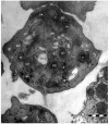
Figure 1
Figure 1
Platelet. Transmission electron microscopy. The platelet cell membrane is encompassing an aliquot of cytoplasm of the thrombocytogenic megakaryocyte, in which different organelles that can be visible (e.g., the Golgi apparatus, GA, mitochondria, m, endoplasmic reticulum, ER), but also the open canalicular system (OCS), alpha granules (αG) and dense granules (δG). The lisosomes with a bundle-like appearance are disposed among the elements of OCS. Randomly dispersed glycogen granules (g) can be observed.
Figure 2
Figure 2
Skin; hair follicle (the outer sheath) and the surrounding dermis. Transmission electron microscopy. Two telocytes (TCs; TC1 and TC2) are located beneath the basement membrane. Within dermis, TCs are presenting cellular prolongations of uneven calibre—telopodes (Tps)—parallel to the basement membrane, and Tps are supplementary branched. Tps are long cellular structures, their entire length being partially intercepted by this plane of section. Supplementary, TC2 (through its Tps) is establishing close contact with a mast cell (MC).
Figure 3
Figure 3
Skin, eccrine sweat gland. Transmission electron microscopy. Neighbouring telocytes (cellular bodies were not encompassed into the microscopic field) are establishing cell contacts by their cellular processes—telopodes (Tps). Moreover, the Tps are branching and enwrapping a nerve ending (n), thus repeating structural positioning motif found in different location of the skin, but also within interstitial space of other organs. Telocytes locations are outside the basal lamina (bm) of the eccrine sweat gland. Above the basal lamina, the basal pole of the clear epithelial secretory cells extensively containing mitochondria (m), ribosomes (r) and elements of the rough endoplasmic reticulum (rER). Aside the clear epithelial secretory cell, two myoepithelial cells (mEp) are visible. Supplementary, the creases of the basal-lateral sides plasma membranes are extensively amplified, creating a labyrinthine structure specific for the clear epithelial secretory cells.
Figure 4
Figure 4
Reticular dermis. Transmission electron microscopy. Telocytes (TCs1-5) and their telopodes (Tps) are surrounding closely a blood vessel. TCs are in the range of molecular contacts to ab luminal surface of the endothelial cells (End) and smooth muscle cells (SMC). Moreover, Tps most probably belonging to other TCs (not encompassed by this plane of section) are concentrically disposed and seem to be involved into an external network of TCs. They are in contact with other types of interstitial cells—lymphocyte (L), macrophages—Mfg. Various packages of collagen fibres (coll) of different thickness are irregularly disposed within reticular dermis.
Figure 5
Figure 5
Dermis. Transmission electron microscopy. A typical ultrastructural aspect of a dermis. Telocytes are displaying the well acknowledged morphology: a small cell body but long, tortuous, and lumpy cellular prolongations—telopodes (Tps). By Tps, TCs are involved in close vicinity with capillaries (cap), and different types of immune cells, e.g., mast cells (MC) and monocytes (Mo). Fragments of Tps (belonging to other TCs outside the plane of section) are apparently randomly disposed, highlighting the acknowledged distribution of TCs in 3-dimensional networks, thus supporting their attributed presumptive roles. End—endothelial cells; N—nerve ending; coll—collagen fibers.

References

    1. Gawaz M., Vogel S. Platelets in Tissue Repair: Control of Apoptosis and Interactions with Regenerative Cells. Blood. 2013;122:2550–2554. doi: 10.1182/blood-2013-05-468694. - DOI - PubMed
    1. Gong T., Liu Y.-T., Fan J. Exosomal Mediators in Sepsis and Inflammatory Organ Injury: Unraveling the Role of Exosomes in Intercellular Crosstalk and Organ Dysfunction. Mil. Med. Res. 2024;11:24. doi: 10.1186/s40779-024-00527-6. - DOI - PMC - PubMed
    1. Wang Q., Liu J., Li R., Wang S., Xu Y., Wang Y., Zhang H., Zhou Y., Zhang X., Chen X., et al. Assessing the Role of Programmed Cell Death Signatures and Related Gene TOP2A in Progression and Prognostic Prediction of Clear Cell Renal Cell Carcinoma. Cancer Cell Int. 2024;24:164. doi: 10.1186/s12935-024-03346-w. - DOI - PMC - PubMed
    1. An N., Chen Z., Zhao P., Yin W. Extracellular Vesicles in Sepsis: Pathogenic Roles, Organ Damage, and Therapeutic Implications. Int. J. Med. Sci. 2023;20:1722–1731. doi: 10.7150/ijms.86832. - DOI - PMC - PubMed
    1. Effah C.Y., Ding X., Drokow E.K., Li X., Tong R., Sun T. Bacteria-Derived Extracellular Vesicles: Endogenous Roles, Therapeutic Potentials and Their Biomimetics for the Treatment and Prevention of Sepsis. Front. Immunol. 2024;15:1296061. doi: 10.3389/fimmu.2024.1296061. - DOI - PMC - PubMed

Publication types

LinkOut - more resources