. 2024 Aug 17;13(16):1373.
doi: 10.3390/cells13161373.

Direct Binding of Synaptopodin 2-Like Protein to Alpha-Actinin Contributes to Actin Bundle Formation in Cardiomyocytes

Affiliations

Direct Binding of Synaptopodin 2-Like Protein to Alpha-Actinin Contributes to Actin Bundle Formation in Cardiomyocytes

Hiroshi Yamada et al. Cells. .

Abstract

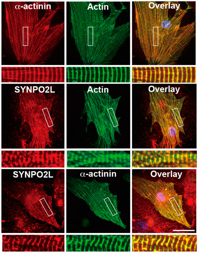
Synaptopodin 2-like protein (SYNPO2L) is localized in the sarcomere of cardiomyocytes and is involved in heart morphogenesis. However, the molecular function of SYNPO2L in the heart is not fully understood. We investigated the interaction of SYNPO2L with sarcomeric α-actinin and actin filaments in cultured mouse cardiomyocytes. Immunofluorescence studies showed that SYNPO2L colocalized with α-actinin and actin filaments at the Z-discs of the sarcomere. Recombinant SYNPO2La or SYNPO2Lb caused a bundling of the actin filaments in the absence of α-actinin and enhanced the α-actinin-dependent formation of actin bundles. In addition, high-speed atomic force microscopy revealed that SYNPO2La directly bound to α-actinin via its globular ends. The interaction between α-actinin and SYNPO2La fixed the movements of the two proteins on the actin filaments. These results strongly suggest that SYNPO2L cooperates with α-actinin during actin bundle formation to facilitate sarcomere formation and maintenance.

Keywords: SYNPO2L; actin; actinin; cardiomyocyte; sarcomere.


Conflict of interest statement

The authors declare no competing interests.

Figures

Figure 1
Figure 1
SYNPO2L, α-actinin, and actin filaments accumulate at the Z-discs of the sarcomere in mouse cardiomyocytes. Double immunofluorescence images of α-actinin and actin filaments (upper panels), SYNPO2L and actin filaments (middle panels) or SYNPO2L and α-actinin (bottom panels) in mouse cardiomyocytes. Areas indicated by rectangles are enlarged in the underneath image. Bar: 30 μm.
Figure 2
Figure 2
SYNPO2La or SYNPO2Lb alters the shape of the α-actinin dimer in vitro. (A) SDS-PAGE of α-actinin, SYNPO2La or SYNPO2Lb. Alpha-actinin (5 μg), SYNPO2La (5 μg), and SYNPO2Lb (10 μg) were analyzed and visualized by SYPRO Orange staining. (BF) Morphological changes of α-actinin dimers in the presence of SYNPO2La or SYNPO2Lb. One micromolar of α-actinin (B), SYNPO2La (C), SYNPO2Lb (D), α-actinin and SYNPO2La (E) or α-actinin and SYNPO2Lb (F) was incubated for 3 h at room temperature. The samples negatively stained and observed by TEM. Scale bar: 100 nm.
Figure 3
Figure 3
SYNPO2La or SYNPO2Lb directly bundles actin filaments and SYNPO2La causes the formation of thick actin bundles. One micromolar α-actinin, SYNPO2La, and SYNPO2Lb were mixed with 4 μM preformed actin filaments and incubated for 3 h at room temperature. The mixture was negatively stained and assessed by TEM at low magnification (AC,GI) or high magnification (DF,JL). Actin filaments alone (A,D). The presence of SYNPO2La or SYNPO2Lb enhanced the formation of thick and long actin bundles. Scale bar: 5 μm in (AC,GI), 300 nm in (DF,JL). (M) Morphometric analysis of actin bundle formation by the indicated proteins. The formation of actin filament bundles was determined by densitometry using Image J analysis of TEM images taken at low magnification (×300). The results were obtained from three independent experiments. * p < 0.05, ** p < 0.01. (N) The diameter of actin bundles formed by the indicated proteins. The diameter of each actin bundle was measured at three different sites of the TEM image taken at high magnification (×20,000). The results were obtained from three independent experiments. ** p < 0.01.
Figure 4
Figure 4
Spatio-temporal high-speed AFM observations of SYNPO2La binding to α-actinin. (A) SYNPO2La (11 nM) formed various shapes and sometimes had a globular shape. (B) Alpha-actinin (50 nM) formed dimers that had a globular shape at both ends. (C) Time-lapsed high-speed AFM images of SYNPO2La (11 nM). (D) Time-lapsed high-speed AFM images of α-actinin (50 nM)/SYNPO2La (28 nM). Scale bar in (AD): 20 nm.
Figure 5
Figure 5
Spatio-temporal characteristics of actin-filament crosslinking mediated by α-actinin and/or SYNPO2La. (A) High-speed AFM image of α-actinin (140 nM)/actin filaments. Alpha-actinin caused the crosslinking of adjacent actin filaments into a ladder-like structure (indicated by the white arrowheads) and bound to the actin filament (indicated by the red arrowheads). Scale bar: 50 nm. (B) High-speed AFM image of SYNPO2La (120 nM)/actin filaments. SYNPO2La forms aggregates, which are bound to and crosslinked actin filaments. Scale bar: 50 nm. (C) High-speed AFM image of α-actinin (140 nM)/SYNPO2La (200 nM)/actin filaments. Globular, rod-like or complex shapes were observed on the actin filaments. Scale bar: 50 nm. (D) Time-lapsed high-speed AFM images of α-actinin/SYNPO2La/actin filaments. The α-actinin/SYNPO2La complex that was bound to actin filaments rarely moved along bundled actin filaments (indicated by the white arrowheads). Scale bar: 50 nm.

References

    1. Sjöblom B., Salmazo A., Djinović-Carugo K. Alpha-actinin structure and regulation. Cell Mol. Life Sci. 2008;65:2688–2701. doi: 10.1007/s00018-008-8080-8. - DOI - PMC - PubMed
    1. Chalovich J.M., Schroeter M.M. Synaptopodin family of natively unfolded, actin binding proteins: Physical properties and potential biological functions. Biophys. Rev. 2010;2:181–189. doi: 10.1007/s12551-010-0040-5. - DOI - PMC - PubMed
    1. Beqqali A., Monshouwer-Kloots J., Monteiro R., Welling M., Bakkers J., Ehler E., Verkleij A., Mummery C., Passier R. CHAP is a newly identified Z-disc protein essential for heart and skeletal muscle function. J. Cell Sci. 2010;123:1141–1150. doi: 10.1242/jcs.063859. - DOI - PubMed
    1. Van Eldik W., den Adel B., Monshouwer-Kloots J., Salvatori D., Maas S., van der Made I., Creemers E.E., Frank D., Frey N., Boontje N., et al. Z-disc protein CHAPb induces cardiomyopathy and contractile dysfunction in the postnatal heart. PLoS ONE. 2017;12:e0189139. doi: 10.1371/journal.pone.0189139. - DOI - PMC - PubMed
    1. La T.M., Tachibana H., Li S.A., Abe T., Seiriki S., Nagaoka H., Takashima E., Takeda T., Ogawa D., Makino S.I., et al. Dynamin 1 is important for microtubule organization and stabilization in glomerular podocytes. FASEB J. 2020;34:16449–16463. doi: 10.1096/fj.202001240RR. - DOI - PubMed

Publication types

LinkOut - more resources