Review
. 2024 Aug 22;13(16):1400.
doi: 10.3390/cells13161400.

New Insights into D-Aspartate Signaling in Testicular Activity

Affiliations
Review

New Insights into D-Aspartate Signaling in Testicular Activity

Sara Falvo et al. Cells. .

Abstract

D-aspartate (D-Asp) is an amino acid found in high concentrations in the testis and pituitary gland. Increasing evidence suggests that D-Asp promotes spermatogenesis by activating testosterone production in the Leydig cells via LH release from the pituitary gland. In vitro studies indicate that D-Asp may also influence steroidogenesis and spermatogenesis through autocrine and paracrine signals. D-Asp enhances StAR and steroidogenic enzyme expressions, facilitating testicular cell proliferation via the GluR/ERK1/2 pathway. Moreover, it supports spermatogenesis by enhancing the mitochondrial function in spermatocytes, aiding in the metabolic shift during meiosis. Enhanced mitochondrial function, along with improved MAM stability and reduced ER stress, has been observed in Leydig and Sertoli cells treated with D-Asp, indicating potential benefits in steroidogenesis and spermatogenesis efficiency. Conversely, D-Asp exerts a notable anti-apoptotic effect in the testis via the AMPAR/AKT pathway, potentially mediated by antioxidant enzyme modulation to mitigate testicular oxidative stress. This review lays the groundwork for future investigations into the molecules promoting spermatogenesis by stimulating endogenous testosterone biosynthesis, with D-amino acids emerging as promising candidates.

Keywords: AKT; D-amino acids; D-aspartate; ERK1/2; MAMs; mitochondria; reproduction; spermatogenesis; steroidogenesis; testis.


Conflict of interest statement

The authors declare no conflicts of interest.

Figures
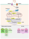
Figure 1
Figure 1
Effects of D-Asp on Leydig cell function. In Leydig cells, D-Asp promotes the phosphorylation of ERK via NMDAR/AMPAR/cAMP. ERK activates gene transcription/protein expression of StAR and steroidogenic enzymes (P450scc, 3β-HSD, 17β-HSD, 5α-red), resulting in the biosynthesis of androgen hormones (testosterone; dihydrotestosterone, DHT). D-Asp also enhances the expression of proteins involved in mitochondrial biogenesis (NRF1, TFAM), fusion (MFN1, MFN2, OPA1), and mass (TOMM20), and reduces the expression of DRP1, a marker of mitochondrial fission. Finally, the amino acid induces an increase in the expression of proteins involved in MAM structure stabilization (ATAD3A, FACL4, SOAT1, VDAC, GRP75), and a reduction in the expression of GRP78, which is indicative of a decrease in ER stress.
Figure 2
Figure 2
D-Asp modulates spermatogenesis through the GluR/ERK1/2/AKT pathways. D-Asp activates the ERK/AKT pathway via GluRs (NMDAR and AMPAR) in testicular germ cells (spermatogonia and spermatocytes) and somatic cells (Leydig and Sertoli cells). ERK activates the expression of proteins involved in mitotic and meiotic processes (PCNA, AuroraB, pH3, SYCP3). D-Asp, via the AMPAR-dependent molecular pathway, elicits an increase in PREP protein by mediating calcium signaling; in spermatogonia the amino acid upregulates the expression of DAAM protein and mediates its transfer into the nucleus. Finally, D-Asp increases the levels of antioxidant enzymes (SOD, CAT), and induces a decrease in TBARS and MDA, resulting in a reduction in oxidative status; by promoting AKT phosphorylation, the amino acid inhibits the apoptotic process, as evidenced by decreased expression levels of Cyt c and caspase-3 and a decreased Bax/Bcl2 ratio.
Figure 3
Figure 3
Effects of D-Asp on Sertoli cell function. D-Asp influences the activity of TM4 Sertoli cells via the ERK/AKT pathway. The phosphorylation of ERK induces an increase in the expression of AR, and vice versa, the phosphorylation of AKT provokes a reduction in the expression of apoptotic markers (Cyt c and the Bax/Bcl2 ratio). Furthermore, D-Asp enhances the MMP and the expression of OXPHOS and proteins involved in mitochondrial biogenesis (PGC1α, NRF1, and TFAM), mass (TOMM20), fission (DRP1), and fusion (MFN1 and MFN2). D-Asp promotes an increase in the expression of the proteins involved in MAM structure stabilization (GRP75, VDAC, FACL4, SOAT1) and a reduction in GRP78 expression, which is indicative of a decrease in ER stress. Finally, a reduction of oxidative stress, as indicated by decreased MDA levels and increased SOD1/2 and CAT expression levels, was described in D-Asp-treated TM4 cells.

References

    1. D’Aniello A. D-Aspartic acid: An endogenous amino acid with an important neuroendocrine role. Brain Res. Rev. 2007;53:215–234. doi: 10.1016/j.brainresrev.2006.08.005. - DOI - PubMed
    1. Hamase K. Sensitive two-dimensional determination of small amounts of d-amino acids in mammals and the study on their functions. Chem. Pharm. Bull. 2007;55:503–510. doi: 10.1248/cpb.55.503. - DOI - PubMed
    1. Hamase K., Morikawa A., Etoh S., Tojo Y., Miyoshi Y., Zaitsu K. Analysis of small amounts of d-amino acids and the study of their physiological functions in mammals. Anal. Sci. 2009;25:961–968. doi: 10.2116/analsci.25.961. - DOI - PubMed
    1. Di Fiore M.M., Santillo A., Chieffi Baccari G. Current knowledge of d-aspartate in glandular tissues. Amino Acids. 2014;46:1805–1818. doi: 10.1007/s00726-014-1759-2. - DOI - PubMed
    1. Karakawa S., Shimbo K., Yamada N., Mizukoshi T., Miyano H., Mita M., Lindner W., Hamase K. Simultaneous analysis of d-alanine, d-aspartic acid, and d-serine using chiral high-performance liquid chromatography-tandem mass spectrometry and its application to the rat plasma and tissues. J. Pharm. Biomed. Anal. 2015;115:123–129. doi: 10.1016/j.jpba.2015.05.024. - DOI - PubMed

LinkOut - more resources