Review
. 2024 Jul 29;22(8):349.
doi: 10.3390/md22080349.

Functional Diversity and Engineering of the Adenylation Domains in Nonribosomal Peptide Synthetases

Affiliations
Review

Functional Diversity and Engineering of the Adenylation Domains in Nonribosomal Peptide Synthetases

Mengli Zhang et al. Mar Drugs. .

Abstract

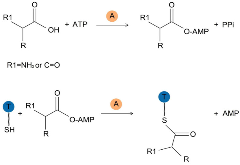
Nonribosomal peptides (NRPs) are biosynthesized by nonribosomal peptide synthetases (NRPSs) and are widely distributed in both terrestrial and marine organisms. Many NRPs and their analogs are biologically active and serve as therapeutic agents. The adenylation (A) domain is a key catalytic domain that primarily controls the sequence of a product during the assembling of NRPs and thus plays a predominant role in the structural diversity of NRPs. Engineering of the A domain to alter substrate specificity is a potential strategy for obtaining novel NRPs for pharmaceutical studies. On the basis of introducing the catalytic mechanism and multiple functions of the A domains, this article systematically describes several representative NRPS engineering strategies targeting the A domain, including mutagenesis of substrate-specificity codes, substitution of condensation-adenylation bidomains, the entire A domain or its subdomains, domain insertion, and whole-module rearrangements.

Keywords: adenylation domain in nonribosomal peptide synthetase; interrupted adenylation domain with methylase activity; nonribosomal peptide synthetase engineering targeting the adenylation domain; substrate-specificity codes of the adenylation domain.


Conflict of interest statement

The authors declare no conflicts of interest.

Figures

Figure 1
Figure 1
The typical domain arrangement of NRPS. Adenylation (A) domain, thiolation (T) domain, condensation (C) domain, and thioesterase (TE) domain are represented in orange, blue, green, and pink circles, respectively. Note: The module illustrated in the figure refers to a unit that comprises the three core domains C, A, and T (the initiation module, shown on the left, consists of A and T domains only, while the termination module, illustrated on the right, contains C, A, T and TE domains).
Figure 2
Figure 2
Activation and loading of substrate catalyzed by the A domain. Initially, the A domain activates the acyl monomer: substrate reacts with adenosine 5′-triphosphate (ATP) to generate the acyl-adenosine monophosphate (AMP) intermediate and inorganic pyrophosphate (PPi). Subsequently, the aminoacyl-AMP undergoes nucleophilic attack by the thiol group located at the terminus of the 4′-phosphopantetheine arm of the downstream thiolation (T) domain, leading to the formation of a thioester-bound aminoacyl-S-T domain by linking to the thiol of the phosphopantetheine arm of T domain, followed by the release of AMP.
Figure 3
Figure 3
Composition of some interrupted A domains: In KtzH, an interruption occurs between a8 and a9 codes of the A domain in module 4, where an O-methylase domain is inserted [55]. In TioS, interruptions occur between a8 and a9 codes of the A domain in both module 3 and module 4, with the insertion of an N-methylase domain [53]. In TioN, an interruption occurs between a2 and a3 codes of the A domain, with the insertion of an S-methylase domain [50]. In MarQ, an interruption occurs between a2 and a3 codes of the A domain, with the insertion of an S-methylase domain [57]. Mb: main-chain methylase domain (yellow); Ms: side-chain methylase domain (light green).
Figure 4
Figure 4
Two typical cases of A domain substitution. (A) The A domain in Bacillus subtilis SrfA-C, which recognizes Leu, is replaced with an A domain that recognizes Cys. (B) The A domain of the second module of PvdD that recognizes L-Thr along with the linker region is replaced with a randomly selected one from nine different types of A domains that recognize other substrates and their corresponding linker regions. Among these, substitutions of six exchanged A domains labeled with dashed circles achieved higher yields. Abbreviations: aad: δ-L-α-aminoadipyl residue, fhOrn: N5-formyl-N5-hydroxyornithine residue.
Figure 5
Figure 5
The cartoon representation of the overall structure of GrsA (PDB ID: 1AMU), phenylalanine-selective A domain of gramicidin synthetase 1, showing the subdomain and core domain of the A domain, which are represented in yellow and cyan, respectively.
Figure 6
Figure 6
The second A domain of EndA that recognizes L-Thr. The FSD responsible for substrate recognition in this domain is replaced with the FSD from EndC that recognizes Ser, enabling the second A domain of EndA to recognize Ser. Abbreviation: Hpg: hydroxyphenylglycine.
Figure 7
Figure 7
Several typical cases of sequence removal and insertion in A domains. (A) The wild-type A domain of KtzH is naturally interrupted. The methylase activity of this domain is abolished after its interrupted Ms domain is removed. (B) Replacing the interrupted Ms domain in KtzH with the interrupted M domain from TioN yields an A domain exhibiting both methylation and adenylation functions; inserting the interrupted M domains from KtzH and TioS into the uninterrupted A domain of Ecm6 results in the generation of corresponding A domains with dual functions of methylation and adenylation.
Figure 8
Figure 8
The domain arrangement and overall structure of SrfA-C. (A) The domain arrangement of the SrfA-C module: C domain, A domain, T domain, TE domain, and linker regions that connect these domains are represented as green, orange, blue, pink, and gray, respectively. (B) The surface representation of the overall structure of SrfA-C (PDB ID: 2VSQ), as well as the colors of the domains and linker regions, are identical to that in (A).
Figure 9
Figure 9
Two typical cases of whole-module rearrangement. (A) Deleting the second module in SrfA-A and connecting modules 1 and 3 resulted in the production of a hexapeptide lacking a Leu residue. (B) By the insertion of a composite module composed of the T-E bidomain from module 4 and the C-A bidomain from module 5 in BpsB into modules 4 and 5 of BpsB, leading to the production of a novel octapeptide with three Hpg residues. Abbreviation: Hpg: hydroxyphenylglycine.

References

    1. Izore T., Cryle M.J. The many faces and important roles of protein-protein interactions during non-ribosomal peptide synthesis. Nat. Prod. Rep. 2018;35:1120–1139. doi: 10.1039/C8NP00038G. - DOI - PubMed
    1. Payne J.A.E., Schoppet M., Hansen M.H., Cryle M.J. Diversity of nature’s assembly lines recent discoveries in non-ribosomal peptide synthesis. Mol. Biosyst. 2017;13:9–22. doi: 10.1039/C6MB00675B. - DOI - PubMed
    1. Miller B.R., Gulick A.M. Structural Biology of Nonribosomal Peptide Synthetases. Methods Mol. Biol. 2016;1401:3–29. doi: 10.1007/978-1-4939-3375-4_1. - DOI - PMC - PubMed
    1. Walsh C.T., Brien R.V.O., Khosla C. Nonproteinogenic Amino Acid Building Blocks for Nonribosomal Peptide and Hybrid Polyketide Scaffolds. Angew. Chem.-Int. Ed. 2013;52:7098–7124. doi: 10.1002/anie.201208344. - DOI - PMC - PubMed
    1. Duckworth B.P., Wilson D.J., Aldrich C.C. Measurement of Nonribosomal Peptide Synthetase Adenylation Domain Activity Using a Continuous Hydroxylamine Release Assay. Methods Mol. Biol. 2016;1401:53–61. doi: 10.1007/978-1-4939-3375-4_3. - DOI - PMC - PubMed

Substances

LinkOut - more resources