. 2024 Aug 18;14(8):456.
doi: 10.3390/metabo14080456.

ATP, the 31P Spectral Modulus, and Metabolism

Affiliations

ATP, the 31P Spectral Modulus, and Metabolism

Jack V Greiner et al. Metabolites. .

Abstract

Adenosine triphosphate (ATP) has a high intracellular millimolar concentration (ca. 2.4 mM) throughout the phylogenetic spectrum of eukaryotes, archaea, and prokaryotes. In addition, the function of ATP as a hydrotrope in the prevention of protein aggregation and maintenance of protein solubilization is essential to cellular, tissue, and organ homeostasis. The 31P spectral modulus (PSM) is a measure of the health status of cell, tissue, and organ systems, as well as of ATP, and it is based on in vivo 31P nuclear magnetic resonance (31P NMR) spectra. The PSM is calculated by dividing the area of the 31P NMR integral curve representing the high-energy phosphates by that of the low-energy phosphates. Unlike the difficulties encountered in measuring organophosphates such as ATP or any other phosphorylated metabolites in a conventional 31P NMR spectrum or in processed tissue samples, in vivo PSM measurements are possible with NMR surface-coil technology. The PSM does not rely on the resolution of individual metabolite signals but uses the total area derived from each of the NMR integral curves of the above-described spectral regions. Calculation is based on a simple ratio of the high- and low-energy phosphate bands, which are conveniently arranged in the high- and low-field portions of the 31P NMR spectrum. In practice, there is essentially no signal overlap between these two regions, with the dividing point being ca. -3 δ. ATP is the principal contributor to the maintenance of an elevated PSM that is typically observed in healthy systems. The purpose of this study is to demonstrate that (1) in general, the higher the metabolic activity, the higher the 31P spectral modulus, and (2) the modulus calculation does not require highly resolved 31P spectral signals and thus can even be used with reduced signal-to-noise spectra such as those detected as a result of in vivo analyses or those that may be obtained during a clinical MRI examination. With increasing metabolic stress or maturation of metabolic disease in cells, tissues, or organ systems, the PSM index declines; alternatively, with decreasing stress or resolution of disease states, the PSM increases. The PSM can serve to monitor normal homeostasis as a diagnostic tool and may be used to monitor disease processes with and without interventional treatment.

Keywords: 31P nuclear magnetic resonance; 31P spectral modulus; ATP; hydrotrope; lens; protein aggregation.


Conflict of interest statement

The authors declare no conflicts of interest.

Figures

Figure 1
Figure 1
Atomic molecular structure of adenosine triphosphate (ATP) [9] depicting the adenine moiety, the adenosine moiety, comprising purine adenine and the sugar ribose, and the triphosphate residue connected by the sugar ribose. γ, β, and α phosphorus moieties comprising the triphosphate residue are depicted.
Figure 2
Figure 2
Phosphorus-31 nuclear magnetic resonance (31P NMR) spectrum of an intact Tegu lizard crystalline lens with the γ-, α-, and β-resonance signals of ATP (adenosine triphosphate). The δ (ppm) scale follows the shift convention of the International Union for Pure and Applied Chemistry (IUPAC) and is referenced relative to the resonance position of 85% phosphorus acid [17,18], αGP, alpha-glycerophosphate signal part of the sugar phosphate groups; Pi, inorganic orthophosphate; GPC, glycerolphosphorylcholine; ADP, adenosine diphosphate; DN, the dinucleotides; NS, nucleoside sugar phosphates. The identity of the spectral shift positions assigned to each organophosphate in the above spectrum are recognized in Methods in Enzymology [4].
Figure 3
Figure 3
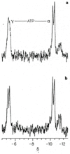
Phosphorus-31 nuclear magnetic resonance (31P NMR) spectrum of the γ- and α-group resonance signals of the tripolyphosphate residue of ATP from an ex vivo intact canine crystalline lens [19]. The identity of the spectral shift positions assigned to the ATP γ- and α-group phosphates in the above spectra are recognized in Methods in Enzymology [4]. The signal splitting phenomenon with doublet formation, as illustrated above, has been well described [19]. (a) Control demonstrates broadened phosphate signal lines of the ATP γ-group doublet due to rapid proton exchange of this weak-acid ATP phosphate group with the protons of the surrounding interstitial water solvent. (b) Ex vivo 31P NMR spectrum from an intact canine lens incubated for 3.2 h in D2O, with the D2O-narrowed signal lines of the ATP γ-group doublet resulting from exchange substitution of the ATP γ-group protons associated with the surrounding hydrogen atoms of the water molecules with the deuterium atom now present in the interstitial water solvent. The γ-phosphate signal narrows because, unlike the protons of the water molecule, the deuterium nucleus spin-couples poorly with the 31P phosphate nucleus. Note that both the control and deuterium-incubated spectra were obtained using the same lens and identical NMR instrumental settings. Filter time constant used introduced 1 Hz line broadening to the spectra. Ordinarily, filter time constants introducing 10 Hz or greater line broadening are used in intact-tissue 31P spectra. Such filtering usually masks the fine structure of ATP [21].
Figure 4
Figure 4
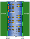
Schematic representation depicting the relationship between adjacent intracellular protein molecules, hydrotropic ATP molecules, and the interfacial space between the protein surfaces [9]. Interstitial water fills the interfacial space (blue), bounded by two adjacent proteins (vertical green lines). The hydrophobic regions of the adjacent proteins (dark green portions of the vertical green lines) interact with the ATP adenine residues (orange). The ATP ribose sugar residue (short wavy line) connects the hydrophobic adenine moiety with the hydrophilic ATP α-, β-, and γ-triphosphate residues (yellow circles). The triphosphate chain residue extends into the interfacial space.
Figure 5
Figure 5
Ex vivo phosphorus-31 nuclear magnetic resonance spectrum and integral curve of a human cornea [26]: (a) sugar phosphate resonance band, (b) inorganic orthophosphate signal, (c) glycerol 3-phosphorylcholine signal, (d) γ-group phosphate resonance of ATP, (e) α-group phosphate resonance of ATP, and (f) β-group phosphate resonance of ATP. The vertical broken line divides low-energy (left) and high-energy spectral regions (right). The quantity of organophosphate beneath the integral curve (g) to the left of the broken line corresponds to the integrated signal area of low-energy phosphates, and the quantity under the integral curve (g) to the right of the vertical broken line corresponds to the integrated signal area of high-energy phosphates. The inflection point is at the intersection of the vertical broken line and the integral curve.

References

    1. Hoult D.I., Busby S.J., Gadian D.G., Radda G.K., Richards R.E., Seeley P.J. Observation of tissue metabolites using 31P nuclear magnetic resonance. Nature. 1974;252:285–287. doi: 10.1038/252285a0. - DOI - PubMed
    1. Bárány M., Bárány K., Burt C.T., Glonek T., Myers T.C. Structural changes in myosin during contraction and the state of ATP in the intact frog muscle. J. Supramol. Struct. 1975;3:125–140. doi: 10.1002/jss.400030205. - DOI - PubMed
    1. Glonek T. Applications of 31P NMR to biological system with emphasis on intact tissue determinations. In: Stec W.J., editor. Phosphorus Chemistry Directed towards Biology: Lectures Presented at the International Symposium Phosphorus Chemistry Directed towards Biology, Burzenin, Poland, 25–28 September 1979. Pergamon Press; Oxford, UK: 1980. pp. 157–174.
    1. Bárány M., Glonek T. Phosphorus-31 nuclear magnetic resonance of contractile systems. Methods Enzymol. 1982;85B:624–676. - PubMed
    1. Greiner J.V., Kopp S.J., Sanders D.R., Glonek T. Organophosphates of the crystalline lens: A nuclear magnetic resonance spectroscopic study. Investig. Ophthalmol. Vis. Sci. 1981;21:700–713. - PubMed

LinkOut - more resources