Case Reports
. 2024 Aug 20;9(8):186.
doi: 10.3390/tropicalmed9080186.

Bartonella Neuroretinitis with Initial Seronegativity and an Absent Macular Star: A Case Report and Literature Review

Affiliations
Case Reports

Bartonella Neuroretinitis with Initial Seronegativity and an Absent Macular Star: A Case Report and Literature Review

Jason Timothy Pan et al. Trop Med Infect Dis. .

Abstract

Cat-scratch disease (CSD) is an infectious disease caused by Bartonella henselae, presenting with fever and lymphadenopathy following contact with felines. The ocular manifestations include neuroretinitis, characterised by optic nerve swelling and a macular star. Case Presentation: We discuss a case of neuroretinitis that presented atypically, without a macular star. There was an initial suspicion of Bartonella, but the serology was negative. Our patient was eventually empirically treated for infective neuroretinitis based on a positive contact history (recently scratched by one of his three pet cats). There was progression to a macular star upon serial dilated fundus examination, and the repeated serology one week after symptom onset showed rising titres, supporting a diagnosis of CSD. Conclusions: A judicious review of systems, repeat assays, serial dilated fundus examination, and early ophthalmic evaluation are useful in cases of suspected neuroretinitis, remaining an important differential in the evaluation of sudden-onset painless vision loss and unilateral disc swelling.

Keywords: Bartonella henselae; cat-scratch disease; neuroretinitis.


Conflict of interest statement

The authors have no competing interests to declare.

Figures
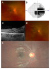
Figure 1
Figure 1
(A) From left to right, fundus photograph of the patient’s right eye at first presentation reveals hyperaemic optic disc swelling with macular oedema covering two disc diameters. (B) Static automatic perimetry shows a centrocecal scotoma in the right eye. (C) Optical coherence tomography of the right macula shows suggestion of intraretinal and subretinal fluid extending from the optic nerve and the presence of hard exudates in Henle’s layer. (D) Fundus photograph showing progression to the formation of a full macular star over the course of 1 week. (E) Fundus photograph after 2 weeks of treatment, showing resolution of optic disc and macular oedema, with residual hard exudates in the configuration of a resolving macular star.

References

    1. Abdelhakim A., Rasool N. Neuroretinitis: A review. Curr. Opin. Ophthalmol. 2018;29:514–519. doi: 10.1097/ICU.0000000000000527. - DOI - PubMed
    1. Joo T., Kim T.G., Moon S.W. Incidental multiple myeloma in a patient with neuroretinitis: A case report. BMC Ophthalmol. 2023;23:495. doi: 10.1186/s12886-023-03220-9. - DOI - PMC - PubMed
    1. Kahloun R., Khairallah-Ksiaa I., Abroug N., Mahmoud A., Ben Yahia S., Zaouali S., Jelliti B., Khairallah M. Final Diagnosis in Patients Referred with a Diagnosis of Neuroretinitis. Neuroophthalmology. 2015;39:266–270. doi: 10.3109/01658107.2015.1092561. - DOI - PMC - PubMed
    1. Nasirudeen A.M., Thong M.L. Prevalence of Bartonella henselae immunoglobulin G antibodies in Singaporean cats. Pediatr. Infect. Dis. J. 1999;18:276–278. doi: 10.1097/00006454-199903000-00014. - DOI - PubMed
    1. Lam J.S., Sanjay S. Lyme neuroretinitis in Singapore: A diagnostic dilemma. Ann. Acad. Med. Singap. 2012;41:178–179. doi: 10.47102/annals-acadmedsg.V41N4p178. - DOI - PubMed

Publication types

LinkOut - more resources