. 2022 Feb;1(2):157-173.
doi: 10.1038/s44161-022-00018-8. Epub 2022 Feb 17.

Extensive identification of genes involved in congenital and structural heart disorders and cardiomyopathy

Nadine Spielmann  1 Gregor Miller  1 Tudor I Oprea  2   3   4 Chih-Wei Hsu  5 Gisela Fobo  1 Goar Frishman  1 Corinna Montrone  1 Hamed Haseli Mashhadi  6 Jeremy Mason  6 Violeta Munoz Fuentes  6 Stefanie Leuchtenberger  1 Andreas Ruepp  1 Matias Wagner  7 Dominik S Westphal  7   8 Cordula Wolf  9   10 Agnes Görlach  11   12 Adrián Sanz-Moreno  1 Yi-Li Cho  1 Raffaele Teperino  1 Stefan Brandmaier  13   14 Sapna Sharma  13   14 Isabella Rikarda Galter  1 Manuela A Östereicher  1 Lilly Zapf  1 Philipp Mayer-Kuckuk  1 Jan Rozman  14   15 Lydia Teboul  16 Rosie K A Bunton-Stasyshyn  16 Heather Cater  16 Michelle Stewart  16 Skevoulla Christou  16 Henrik Westerberg  16 Amelia M Willett  17 Janine M Wotton  17 Willson B Roper  17 Audrey E Christiansen  5 Christopher S Ward  5 Jason D Heaney  5 Corey L Reynolds  5 Jan Prochazka  15 Lynette Bower  18 David Clary  18 Mohammed Selloum  19 Ghina Bou About  19 Olivia Wendling  19 Hugues Jacobs  19 Sophie Leblanc  19 Hamid Meziane  19 Tania Sorg  19 Enrique Audain  20   21 Arthur Gilly  22 Nigel W Rayner  22   23   24 IMPC consortiumGenomics England Research ConsortiumMarc-Phillip Hitz  20   21   25 Eleftheria Zeggini  22   26 Eckhard Wolf  27 Radislav Sedlacek  15 Steven A Murray  17 Karen L Svenson  17 Robert E Braun  17 Jaqueline K White  17 Lois Kelsey  28   29 Xiang Gao  30 Toshihiko Shiroishi  31 Ying Xu  32 Je Kyung Seong  33 Fabio Mammano  34 Glauco P Tocchini-Valentini  34 Arthur L Beaudet  5 Terrence F Meehan  6 Helen Parkinson  6 Damian Smedley  35 Ann-Marie Mallon  16 Sara E Wells  16 Harald Grallert  13   14 Wolfgang Wurst  36   37   38   39 Susan Marschall  1 Helmut Fuchs  1 Steve D M Brown  16 Ann M Flenniken  28   29 Lauryl M J Nutter  28   40 Colin McKerlie  29   40 Yann Herault  19   41 K C Kent Lloyd  18   42 Mary E Dickinson  5 Valerie Gailus-Durner  1 Martin Hrabe de Angelis  43   44   45
Collaborators, Affiliations

Extensive identification of genes involved in congenital and structural heart disorders and cardiomyopathy

Nadine Spielmann et al. Nat Cardiovasc Res. 2022 Feb.

Erratum in

  • Publisher Correction: Extensive identification of genes involved in congenital and structural heart disorders and cardiomyopathy.
    Spielmann N, Miller G, Oprea TI, Hsu CW, Fobo G, Frishman G, Montrone C, Haseli Mashhadi H, Mason J, Munoz Fuentes V, Leuchtenberger S, Ruepp A, Wagner M, Westphal DS, Wolf C, Görlach A, Sanz-Moreno A, Cho YL, Teperino R, Brandmaier S, Sharma S, Galter IR, Östereicher MA, Zapf L, Mayer-Kuckuk P, Rozman J, Teboul L, Bunton-Stasyshyn RKA, Cater H, Stewart M, Christou S, Westerberg H, Willett AM, Wotton JM, Roper WB, Christiansen AE, Ward CS, Heaney JD, Reynolds CL, Prochazka J, Bower L, Clary D, Selloum M, Bou About G, Wendling O, Jacobs H, Leblanc S, Meziane H, Sorg T, Audain E, Gilly A, Rayner NW; IMPC consortium; Genomics England Research Consortium; Hitz MP, Zeggini E, Wolf E, Sedlacek R, Murray SA, Svenson KL, Braun RE, White JK, Kelsey L, Gao X, Shiroishi T, Xu Y, Seong JK, Mammano F, Tocchini-Valentini GP, Beaudet AL, Meehan TF, Parkinson H, Smedley D, Mallon AM, Wells SE, Grallert H, Wurst W, Marschall S, Fuchs H, Brown SDM, Flenniken AM, Nutter LMJ, McKerlie C, Herault Y, Lloyd KCK, Dickinson ME, Gailus-Durner V, Hrabe de Angelis M. Spielmann N, et al. Nat Cardiovasc Res. 2022 May;1(5):529-531. doi: 10.1038/s44161-022-00072-2. Nat Cardiovasc Res. 2022. PMID: 40263883 Free PMC article. No abstract available.

Abstract

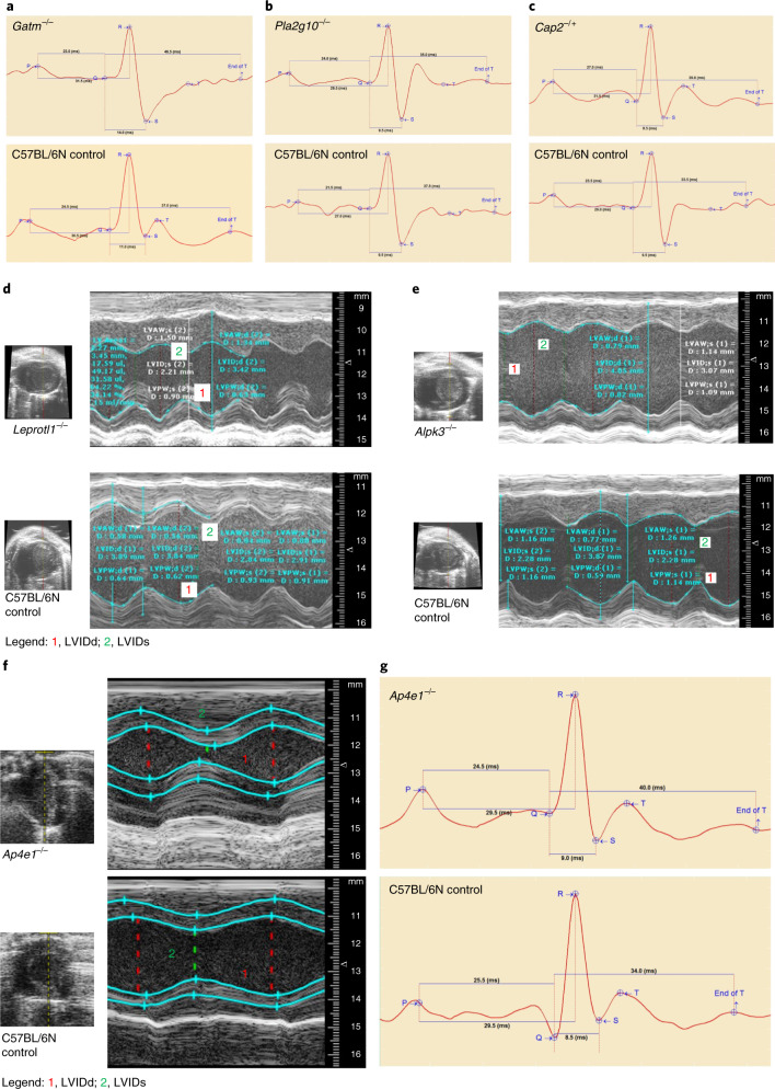
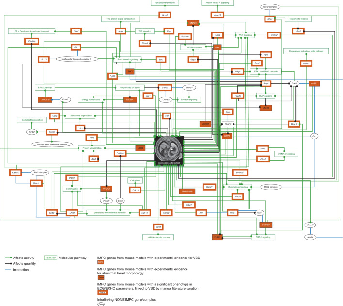
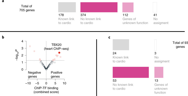
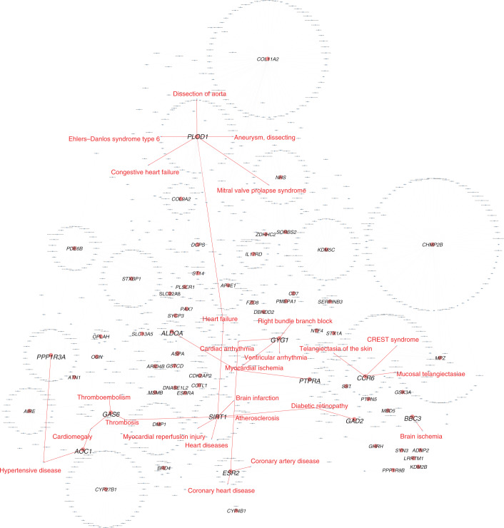
Clinical presentation of congenital heart disease is heterogeneous, making identification of the disease-causing genes and their genetic pathways and mechanisms of action challenging. By using in vivo electrocardiography, transthoracic echocardiography and microcomputed tomography imaging to screen 3,894 single-gene-null mouse lines for structural and functional cardiac abnormalities, here we identify 705 lines with cardiac arrhythmia, myocardial hypertrophy and/or ventricular dilation. Among these 705 genes, 486 have not been previously associated with cardiac dysfunction in humans, and some of them represent variants of unknown relevance (VUR). Mice with mutations in Casz1, Dnajc18, Pde4dip, Rnf38 or Tmem161b genes show developmental cardiac structural abnormalities, with their human orthologs being categorized as VUR. Using UK Biobank data, we validate the importance of the DNAJC18 gene for cardiac homeostasis by showing that its loss of function is associated with altered left ventricular systolic function. Our results identify hundreds of previously unappreciated genes with potential function in congenital heart disease and suggest causal function of five VUR in congenital heart disease.


Conflict of interest statement

T.I.O. has received honoraria or consulted for Abbott, AstraZeneca, Chiron, Genentech, Infinity Pharmaceuticals, Merz Pharmaceuticals, Merck Darmstadt, Mitsubishi Tanabe, Novartis, Ono Pharmaceuticals, Pfizer, Roche, Sanofi and Wyeth. None of these funds were used for this research project.

Figures

Fig. 1
Fig. 1. Candidate genes for cardiac conduction system disease and cardiomyopathy in mice.
Representative electrocardiograms from conscious mutant and control mice with indication of ECG parameters and interval durations. a, Gatm loss (Gatm−/−) caused lower heart rate with prolonged QRS width and QTc and ST interval lengths. b, Pla2g10 loss (Pla2g10−/−) lowered heart rate and prolonged PR, PQ and QTc intervals in female null mice. c, Cap2 depletion (Cap2−/+) induced lower heart rate and lengthy QTc and ST durations in male null mice compared with C57BL/6N controls. Data are presented by Mouse Specifics software. Interval durations are given in milliseconds. M-mode recordings are through a short-axis view tangential to the papillary muscle from representative mutant and control mice. Images show the LVID throughout diastole and systole. d, Leprotl1 depletion (Leprotl1−/−) reduced LV diameters (LVIDs and LVIDd) and increased myocardial wall thickness (LVAWs, LVAWd and LVPWs) with decreased systolic function compared with C57BL/6N controls. e, Alpk3 depletion (Alpk3−/−) increased LV diameters (LVIDs and LVIDd) and decreased systolic function via reduced fractional shortening and ejection fraction, suggesting dilated left ventricle or even dilated cardiomyopathy. The y axis represents the distance (in mm) from the transducer (Vevo 2100); time (in ms) is on the x axis. f, Ap4e1 loss (Ap4e1−/−) caused an impairment of LVIDd and LVIDs and consequently lowered stroke volume. The y axis represents the distance (in mm) from the transducer (Vevo 2100); time (in ms) is on the x axis. g, Representative electrocardiograms from conscious Ap4e1-mutant and C57BL/6N control mice with indication of ECG parameters and interval durations. Ap4e1 loss lowered heart rate and concurrently increased RR interval duration. Data are presented by Mouse Specifics software.
Fig. 2
Fig. 2. VSD network.
Ex situ imaging of the embryo heart in homozygous lethal or homozygous subviable single-gene-knockout mice used to identify structural heart defects, such as VSD. Of the mouse lines studied here, 65% were homozygous knockouts (corresponding to LOF in human) and 35% were heterozygous knockouts. This dataset included 248 of 705 lines (35%). Lines were confirmed to be lethal or subviable; we assessed cardiac development by analyzing three-dimensional micro-CT data obtained from iodine contrast-enhanced micro-CT that provides high spatial resolution of up to 3–14 μm per voxel from embryonic day (E)9.5–E18.5 embryos (http://www.mousephenotype.org/data/embryo). No embryo imaging was performed on viable knockout lines. Embryo data were available for only a small subset of the total knockout genes used for this study. The VSD network included nine genes (Sirt1, Stambp, Casz1, Wfdc2, Tmem161b, Nxn, Dnajc18, Gnao1 and Slc25a) that, when LOF was induced, caused early mortality due to structural heart changes but most notably VSDs in the null mutant mice, experimentally shown by computed tomography microscopy. Five more genes (Zpf503 (human ZNF503), Ubr4, Furin, Shox2 and Smo) were associated with cardiac abnormalities after gene depletion, confirmed by gross morphology data. An additional 45 genes were integrated using their association with cardiac malformations and development of a VSD. Only in vivo ECG and TTE data from young adult mice are available because these mutant lines tested positive for viability; therefore, no embryo screening was performed. The network analysis also identified 13 non-IMPC interacting genes. Two transcription factors, NKX2-5 (Furin, Bmp10, Shox2, Sirt1 and Sspn) and TBX20 (Bmp4, Bmp10, Tfap2b and Casz1), known to be important for early cardiac development, were strongly represented. Furthermore, BMP10, a critical regulator of cardiac growth and chamber maturation, chromatin-modifying (Hdac1 and Smarcb1) genes and genes regulated by processes essential for cardiogenesis, for example, smoothened and WNT signaling (Gnao1, Emilin2, Aldoa, Nxn, Smo, Sufu and Ift81), were strongly represented in the network. Compelling evidence of experimental human or rodent data is from relevant publications, primarily peer-reviewed ‘small-scale experiment’ literature used in the network analysis. ECHO, transthoracic echocardiography; ECG, electrocardiography; NONE, genes not analyzed so far in the IMPC; ER, endoplasmic reticulum.
Fig. 3
Fig. 3. Alignment and enrichment of 705 IMPC knockout genes.
Using Pharos (https://pharos.nih.gov/), a multimodal web interface, we queried alignment for human cardiac disease relevance. We excluded 41 genes that had no human ortholog and/or limited assignment information from this analysis. This analysis indicated that 155 of the 664 mouse–human orthologous genes were previously linked to heart disease. The remaining 509 orthologous genes had not been previously associated with a cardiac condition and likely represent ‘unappreciated candidate genes’ for CHD. This set of 509 genes was further queried for newness using the Online Mendelian Inheritance in Man (OMIM) catalog (https://omim.org/) and Orphanet, a rare disease portal (https://www.orpha.net/) dataset. This secondary analysis confirmed that 486 of 509 genes were unknown candidate genes and the remaining 23 genes have been associated predominantly with pleiotropic neurodevelopmental disorders that include sporadic congenital heart defects or malformations. a, Alignment of 705 IMPC knockout genes using Pharos, OMIM and Orphanet classified them into genes with a previously known link (178 of 705) or unknown link (486 of 705) to CVD or poorly annotated genes (112 of 486); cardio, cardiovascular diseases and/or heart. b, Genes previously identified with abnormal cardiac function in knockout mice (positive genes) showed strong enrichment (P = 0.004; odds ratio, 1.31; combined score, 7.2) for heart-specific targets of the transcription factor TBX20, a critical regulator of heart development–,, associated with human CHD and adult cardiomyopathies,,,, while the 3,189 nonsignificant IMPC knockout genes (negative genes) did not. The ChEA 2016 dataset of publicly available ChIP–seq experiments was used for in silico analysis. Here ‘strong’ enrichment was considered when the following parameters were met: combined score >5 and adjusted P value <0.05; TF, transcription factor. c, Molecular properties of TBX20 target genes showed that 66 of 93 (~70%) have no previous known link to CVD or are poorly annotated genes (13 of 93, ~14%). This observation indicates that, while the role of TBX20 in heart physiology is not completely understood, our dataset sheds light on a previously unknown group of genes with potential relevance for heart development and function.
Fig. 4
Fig. 4. Intersection of mouse and human genes and confirmation of pathogenicity in the heart.
Ortholog matching between our set of 486 non-associated LOF CHD candidate genes and those from two large-scale sequencing studies of patients with CHD and their controls: the US PCGC (https://benchtobassinet.com) and the UK 100KGP (https://www.genomicsengland.co.uk/). a, Intersection analysis with two CHD cohorts, PCGC and 100KGP data revealed that unknown genes from the mouse study emerged in patients with CHD with de novo and LOF variants. Most interestingly, these genes were previously classified as VUR. b, Confirmation of pathogenicity at induction of gene loss in Pde4dip-, Casz1-, Rnf38-, Tmem161b-, Dnajc18- and Rcn3-mutant embryos. These embryonal data show various structural abnormalities of the heart developed during cardiogenesis in knockout mice and confirm causality at gene loss.
Fig. 5
Fig. 5. A bird’s-eye view of the gene–disease association network.
CVD has diverse manifestation. To explore whether our unknown gene candidates also have potential associations with CVD other than CHD, we performed gene–disease enrichment analysis. We performed gene–disease network enrichment analysis for orthologous human–mouse cardiac knockout genes using the automated tool NetworkAnalyst. The gene–disease association network was generated only with the 486 genes that have not yet been associated with cardiac dysfunction in humans. In total, 64 of 486 ‘unknown’ genes were associated with 890 diseases in the enrichment analysis ‘Hairball’ network. Twelve genes were directly related to 25 CVDs, suggesting that they may play a role in human CVDs. Nodes (light red circles), genes from the list of 486 unknown genes; light red circles with larger-font letters, genes associated with CVDs; blue rectangles, diseases; edges colors (red arrows), association of genes with CVDs; light gray arrows, associations of genes with other diseases.
Extended Data Fig. 1
Extended Data Fig. 1. Summary of data gathering and genotype and phenotype information.
a. Schematic overview of the IMPC data gathering. In total, 705 candidate genes with ≥ 1 significant parameter (q-value <0.05) in electrocardiography (ECG) or transthoracic echocardiography (TTE) derivates: ECG n = 424, TTE n = 243, TTE & ECG n = 38 target genes. b: Comprehensive representation of phenotype distribution across 705 genes. Genotype < >phenotype represented by chord graphs. Strength indicated by line thickness (thin lines - low phenotype count). Number of significant phenotypes (called ‘hits’) per gene (q-value <0.05). ECG - left, TTE – middle, both ECG and ECHO - right. Most genes with 1-2 hits, few with ≥ 3 hits. In gene knockouts with abnormal ECG, we observed an abnormal heart rate and inversely correlated RR interval duration in 29% (123/424) of mutant lines. A total of 24% of gene knockouts had QT alterations (102/424), 17% (72/424) in QRS, and 16% (68/424) in ST interval length. In the 243 knockouts with abnormal TTE data, we identified altered fractional shortening and ejection fraction associated with left ventricular dysfunction in 23% (56/243) of gene knockouts. Morphological differences in left ventricular interior diameter (LVID) or left ventricular posterior wall (LVPW) or anterior wall (LVAW) thickness observed in 20% (49/243) of the abnormal TTE knockouts. Half of the gene knockouts with abnormal cardiac phenotypes had a single abnormal phenotype; 49% (207/424) with ECG only and 57% (139/243) with TTE only compared to control animals.
Extended Data Fig. 2
Extended Data Fig. 2. Expression profiles of mouse and human hearts in positive genes (P), CHD-associated genes (CHD) and negative genes (N, reference group).
Gene based expression levels in mouse and human heart tissues performed to evaluate the impact of the size difference between positive and negative gene groups. Expression levels in known CHD genes added at all three stages for reference. Known CHD genes, however, showed significantly increased expression levels compared with positive mouse genes at all three stages. This analysis was further expanded to human heart tissue for genes with a mouse-human orthologue and presented a similar picture. Mean expression levels per groups compared using a Wilcoxon rank-sum test. X-axis denotes the gene groups evaluated; y-axis denotes the log-transformed mean expression in heart. The analysis was stratified by procedure (ECG and TTE) and developmental stages (development, maturation and postnatal). Data shown as mean, minimum, maximum and lower/upper quartiles. a. Expression analysis in mouse heart for ECG genes; b. Expression analysis in mouse heart for TTE genes. Note: RPKM, Reads per kilobase of transcript per Million mapped reads. c. Expression analysis in human heart for ECG genes; d. Expression analysis in human heart for TTE genes. Distribution of the mean expression in mouse heart differences between P and N genes. The negative gene group (higher number of genes) randomly down sampled to generate equal-size subgroups compared to the positive group. P-values denote the probability of observing by chance higher averaged expression level in negative group compared to positive group, in 50,000 permutations. The analysis stratified by procedure (e. ECG and f. TTE) and developmental stages (development, maturation and postnatal. Distribution of the mean expression in human heart differences between P and N genes. g. ECG and h. TTE and developmental stages (development, maturation and postnatal.

References

    1. Watkins, W. S. et al. De novo and recessive forms of congenital heart disease have distinct genetic and phenotypic landscapes. Nat. Commun.10, 4722 (2019). 10.1038/s41467-019-12582-y - DOI - PMC - PubMed
    1. Landstrom, A. P., Dobrev, D. & Wehrens, X. H. T. Calcium signaling and cardiac arrhythmias. Circ. Res.120, 1969–1993 (2017). 10.1161/CIRCRESAHA.117.310083 - DOI - PMC - PubMed
    1. Liu, Y. et al. Global birth prevalence of congenital heart defects 1970–2017: updated systematic review and meta-analysis of 260 studies. Int. J. Epidemiol.48, 455–463 (2019). 10.1093/ije/dyz009 - DOI - PMC - PubMed
    1. Penny, D. J. & Vick, G. W. III Ventricular septal defect. Lancet377, 1103–1112 (2011). 10.1016/S0140-6736(10)61339-6 - DOI - PubMed
    1. Dakkak, W. & Oliver, T. I. Ventricular septal defect. in StatPearlshttps://www.ncbi.nlm.nih.gov/books/NBK470330/ (StatPearls, 2019). - PubMed

LinkOut - more resources