. 2024 Jul;3(7):819-840.
doi: 10.1038/s44161-024-00485-1. Epub 2024 Jul 5.

A common gene signature of the right ventricle in failing rat and human hearts

Affiliations

A common gene signature of the right ventricle in failing rat and human hearts

Liane Jurida et al. Nat Cardiovasc Res. 2024 Jul.

Abstract

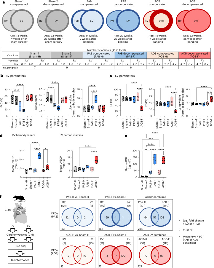
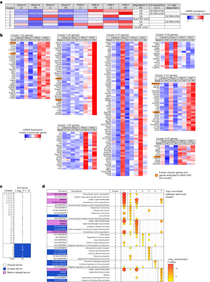
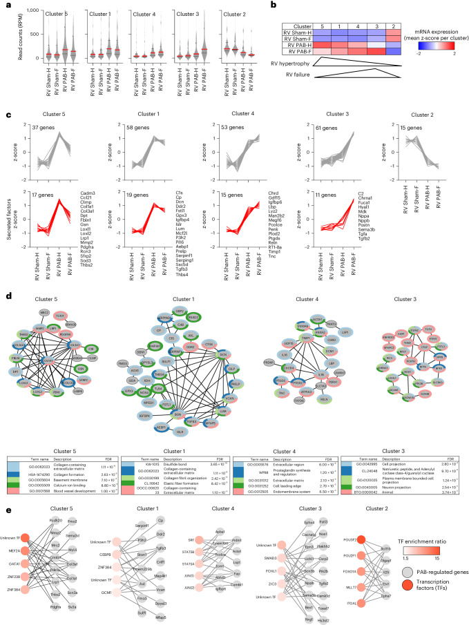
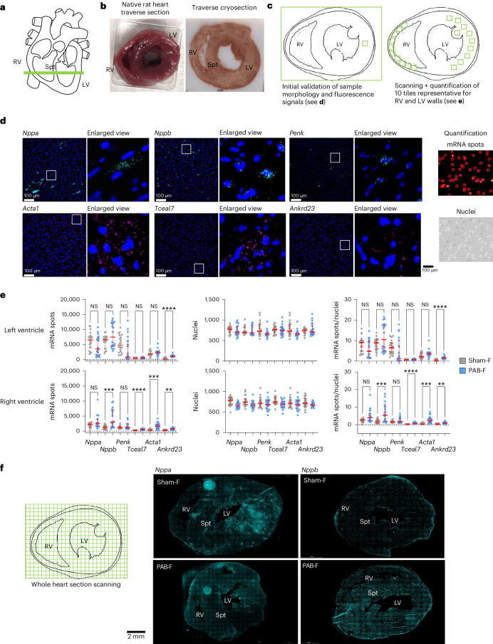
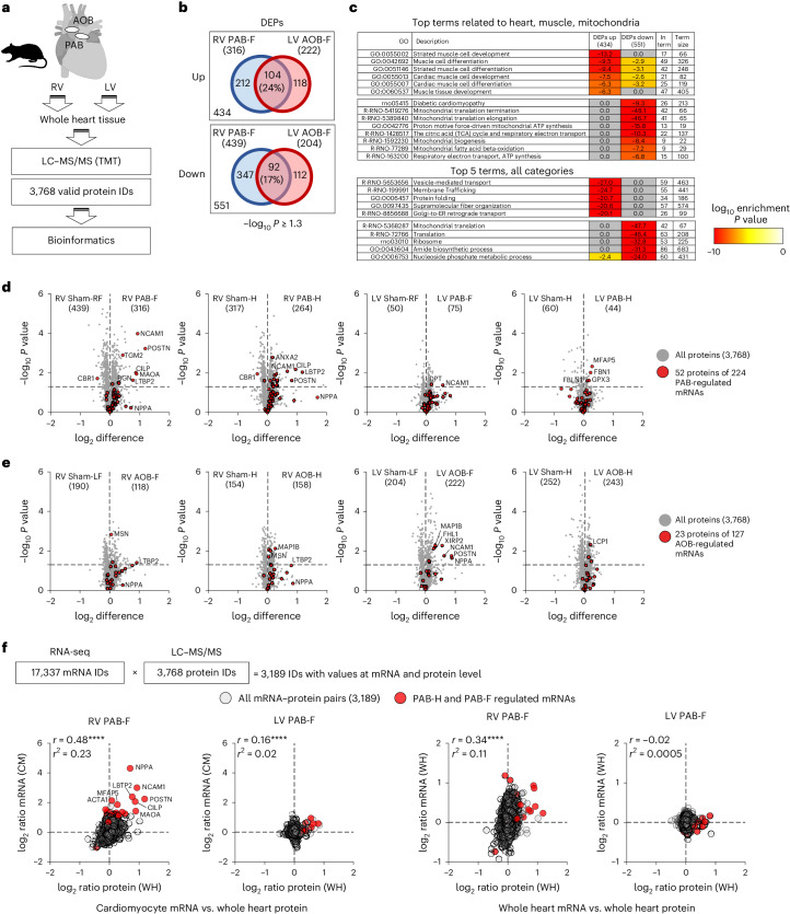
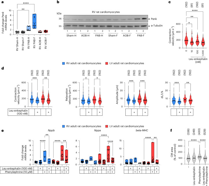
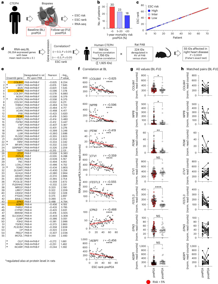
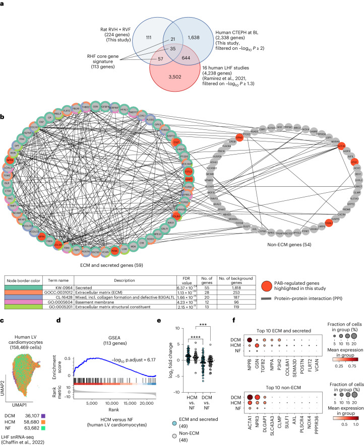
The molecular mechanisms of progressive right heart failure are incompletely understood. In this study, we systematically examined transcriptomic changes occurring over months in isolated cardiomyocytes or whole heart tissues from failing right and left ventricles in rat models of pulmonary artery banding (PAB) or aortic banding (AOB). Detailed bioinformatics analyses resulted in the identification of gene signature, protein and transcription factor networks specific to ventricles and compensated or decompensated disease states. Proteomic and RNA-FISH analyses confirmed PAB-mediated regulation of key genes and revealed spatially heterogeneous mRNA expression in the heart. Intersection of rat PAB-specific gene sets with transcriptome datasets from human patients with chronic thromboembolic pulmonary hypertension (CTEPH) led to the identification of more than 50 genes whose expression levels correlated with the severity of right heart disease, including multiple matrix-regulating and secreted factors. These data define a conserved, differentially regulated genetic network associated with right heart failure in rats and humans.


Conflict of interest statement

The authors declare no competing interests.

Figures

Fig. 1
Fig. 1. Identification of differentially regulated gene sets in cardiomyocytes from rat models of progressive RHF and LHF.
a, Overview of animal study design. PAB or AOB was performed in rats to slowly induce RHF or LHF. Sham-operated animals served as controls. b, Echocardiographic validation of RV function and liver weight of rat models used for CM isolation. c, Echocardiographic validation of LV function and lung weight of rat models used for cardiomyocyte isolation. d, Sham, PAB and AOB surgery was performed in an independent animal cohort. End-diastolic pressure (EDP) was determined by right heart (left graphs) or left heart (right graphs) catheterization in sham controls and after banding at week 14 (compensated stage, PAB-H, AOB-H), at week 29 (PAB decompensated stage, PAB-F) or week 33 (AOB decompensated stage, AOB-F). n = 5 animals per group. e, Plasma BNP levels of compensated and decompensated states in response to PAB or AOB in the rat models from b and c. f, Total RNA from RV and LV cardiomyocytes was isolated, and 17,341 rat genes were analyzed by RNA-seq. Based on two-fold changes, mean read counts in disease conditions of more than 50 and P ≤ 0.01, DEGs were identified in each condition by comparing the PAB or AOB groups to their corresponding sham groups. Venn diagrams indicate overlapping and distinct groups of DEGs for PAB (224 genes) and AOB (127 genes) that were further systematically analyzed in this study. b,c,e,f, n = 6–9 animals per group as shown in a. Box plots in be show data points from all individual animals with means and minimum/maximum values. Asterisks indicate significant changes according to one-way ANOVA (*P ≤ 0.05, **P ≤ 0.01, ***P ≤ 0.001, ****P ≤ 0.0001). RPM, reads per million. Source data
Fig. 2
Fig. 2. Different transcriptomic responses of LVs and RVs to PAB.
a, The mRNA expression values of all 224 PAB-regulated genes (Fig. 1f) from RVs and LVs were hierarchically clustered by k-means. The heatmap shows the averaged z-score-normalized read counts for all genes belonging to each of the seven clusters across all conditions in both LVs and RVs. The prevailing gene regulatory phenotype is indicated in the three right columns. b, Heatmaps of z-scaled mean expression values for all individual 224 genes across the seven clusters and all conditions. Orange colors mark known (Acta1, Cilp, Nppa and Nppb) and new PAB-regulated genes that were highlighted in following analyses or further investigated throughout this study. c, The genes of each cluster were examined for the top 100 overrepresented pathway terms using Metascape (https://www.metascape.org), and multiple Venn analysis was performed to identify overlapping and distinct pathway terms for the gene sets of all seven clusters. Only pathway terms with enrichment −log10 P ≤ 2 were considered. d, Graphical representation of the top five enriched pathways for each cluster along with the corresponding enrichment P values (highlighted by bubble size and color). If below the threshold, values are also included for the other six clusters. GO, Gene Ontology. Source data
Fig. 3
Fig. 3. Identification of gene regulatory networks underlying the transition from RVH to RVF.
a, The mRNA expression values of all 224 PAB-regulated genes from RVs were segregated into five groups by hierarchical k-means clustering. Violin plots show all normalized read counts (reads per million (RPM)), medians (solid red lines) and 1st and 3rd quartiles (dotted red lines). b, z-scaled heatmap of mean mRNA expression for the genes of the five clusters from a. Triangles indicate the overall regulation of clusters 5 and 3, which most specifically characterize the transition from the compensatory to the decompensated states. c, Upper panels: z-scaled expression changes of all individual genes per cluster. Lower panels: subgroups of genes from each cluster (also shown by gene name) encoding secreted factors according to a recent annotation of the secreted proteome. d, Upper part: physical and functional protein networks for all genes (gray nodes) from each cluster shown in c based on STRING database annotations. All PPI categories of STRING (text mining, experiments, databases, co-expression, neighborhood, gene fusion and co-occurrence) and all pathway categories were selected. Depicted are only genes with at least one documented PPI (dark gray edges). All edges had a STRING confidence score greater than 0.4. Lower part: top five enriched pathway terms associated with the genes of each cluster as determined by Cytoscape and the integrated STRING application. Colors of tables and node borders indicate the top five enriched pathway terms associated with each individual gene. FDR indicates the false discovery rate for pathway enrichment. e, All genes of each cluster were examined for their overrepresentation in annotated TF gene sets of the MSigDB (https://www.gsea-msigdb.org/gsea/msigdb). Shown are the top five significantly enriched TFs (reddish nodes) that are connected to their target genes (gray nodes) by gray connecting edges. The complete lists of the top 10 enriched unique TFs and all unique target genes are shown in Supplementary Fig. 2a. The mRNA expression values for these TFs in the rat heart are shown in Supplementary Fig. 2b. Source data
Fig. 4
Fig. 4. Spatial regulation of PAB-regulated genes in the whole rat heart revealed by RNA-FISH.
a, Scheme of the location of traverse sections of the heart used for smRNA-FISH. b, Rat heart samples before and after processing for cryosections. c, Schematic of the strategy for scanning the walls of the RVs and LVs through 10 tiles of equal-sized sections each. d, Representative images of smRNA-FISH performed on 7-µm cryosections. Samples were hybridized with pairs of probes for the indicated transcripts, and mRNAs were visualized by two different fluorophores. Nuclei were stained in parallel with DAPI. White inserts are shown as enlarged view on the right side of each image to demonstrate the spatial distribution of mRNA signals in the dense cardiomyocyte cell layers. Right upper and lower panels demonstrate the automated detection and quantification of mRNA spots (upper image) or nuclei (lower image) of each section by Icy software (version 2.4.2.0) (https://icy.bioimageanalysis.org/). Scale bars indicate 100 µm. Images are representative for one out of two experiments with similar results. e, Rat hearts from controls or PAB-operated animals were obtained at the RHF states (Sham-F, PAB-F) and processed for smRNA-FISH as shown above. The graphs show quantification of mRNA spots, nuclei and mRNA spot signals normalized for cell number of each section according to the nuclei counts. Scatter plots show data from 20 sections derived from two biologically independent experiments. Red lines show medians, and whiskers show 1st and 3rd quartiles. Significant changes were identified by one-way ANOVA; asterisks indicate P values (*P ≤ 0.05, **P ≤ 0.01, ***P ≤ 0.001, ****P ≤ 0.0001). f, Left, schematic strategy for whole heart scans. Right, spatial distribution of Nppa and Nppb mRNA signals across the whole heart before and after PAB. Scale bar, 2,000 µm. Images are representative for one out of two experiments with similar results. NS, not significant; Spt, septum. Source data
Fig. 5
Fig. 5. Proteome analysis of common and differentially regulated factors in the RV and LV of PAB and AOB rat models.
a, Overview of proteomic analyses. Tryptic peptides derived from RV or LV heart tissues of rats subjected to PAB or AOB conditions were labeled with tandem mass tags (TMTs) (n = 8 rats per group). In total, 4,149 protein IDs were identified. Scaled, normalized data were log2 transformed and width normalized. The data matrix was reduced to 3,768 IDs based on 75% (that is, six out of eight) valid values in at least one group. b, Significantly differentially expressed proteins (DEPs) were identified based on pairwise comparisons of AOB or PAB conditions with sham controls using a −log10 P ≥ 1.3. Venn diagrams show the overlap of DEPs in the RV or LV upon PAB or AOB HF conditions. c, All upregulated or downregulated proteins in the PAB or AOB HF conditions as shown in c were pooled and analyzed separately for overrepresented pathway terms related to heart, (cardiac) muscle or mitochondria terms (upper table) or for all functional categories. d,e, Volcano plots show regulation and t-test results of all proteins in PAB or AOB conditions compared to the respective sham controls. Red symbols indicate proteins that matched the differentially expressed mRNAs of PAB (d) or AOB (e) conditions that we identified in Fig. 1f. Numbers in brackets within graphs highlight significantly upregulated or downregulated DEPs (−log10 P ≥ 1.3). f, RNA-seq and proteomic datasets were intersected to identify 3,189 genes with values for both mRNA and protein expression. Graphs show pairwise correlations of PAB-dependent changes for both RV and LV. y axes show changes of mRNA levels from cardiomyocytes (left graphs) or whole heart (right graphs), and x axes show changes of protein levels in whole heart. Red symbols highlight the factors of the principal set of 224 PAB-dependent genes that were regulated at the mRNA level. Pearson correlation coefficient (r) and coefficients of determination (r2) are indicated. Asterisks (****P ≤ 0.0001) indicate P values derived from an F-test to test the null hypothesis that the overall slope is zero. CM, cardiomyocyte; GO, Gene Ontology; WH, whole heart. Source data
Fig. 6
Fig. 6. Validation of PAB-dependent regulation of Penk expression and suppression of cardiomyocyte contractility and hypertrophy by opioid peptides.
a, Penk mRNA expression in RV cardiomyocytes isolated from sham, PAB or AOB conditions was analyzed by RT–qPCR. Box plots show data points from all individual animals with means and minimum/maximum values. Asterisks indicate significant changes according to one-way ANOVA (*P ≤ 0.05, **P ≤ 0.01, ***P ≤ 0.001, ****P ≤ 0.0001). Graphs show all values, including technical duplicates from six biologically independent cardiomyocyte preparations. b, Cell extracts were isolated from pooled RV cardiomyocytes of 6–8 animals per condition and analyzed by western blotting for the expression of Penk. Two independent samples (1 and 2) per condition are shown. Antibodies against tubulin served as a loading control. c,d, Isolated RV or LV cardiomyocytes were paced at 2 Hz. Contraction or relaxation velocity, contraction amplitude and load-free cell shortening (expressed as dL/L (%)) were assessed. c, Dose response of LV cardiomyocytes treated with Leu-enkephalin for 10 min. d, Decreased contractility parameters of RV or LV cardiomyocytes treated with 100 nM Leu-enkephalin for 10 min. Total cell numbers from six independent cardiomyocyte preparations are shown in brackets. e, Isolated RV or LV cardiomyocytes were stimulated with 10 µM phenylephrine for 24 h in the presence or absence of 100 nM Leu-enkephalin. Expression of hypertrophy marker genes was analyzed by RT–qPCR. Changes of mRNA levels were calculated relative to the mean of all untreated controls. Box plots show all data points (including technical replicates) with means and minimum/maximum values obtained from four independent cardiomyocyte preparations. Asterisks indicate significant changes according to one-way ANOVA (*P ≤ 0.05, **P ≤ 0.01, ***P ≤ 0.001, ****P ≤ 0.0001). f, Relative changes of cardiomyocyte area size under the conditions described in e. Total cell numbers from four independent cardiomyocyte preparations are shown in brackets. Solid lines in violin plots in c, d and f show medians; dotted lines show 1st and 3rd quartiles. Significant changes were identified by one-way ANOVA; asterisks indicate P values (*P ≤ 0.05, **P ≤ 0.01, ***P ≤ 0.001, ****P ≤ 0.0001). CM, cardiomyocyte. Source data
Fig. 7
Fig. 7. Rat PAB gene sets are deregulated in patients with CTEPH, and their expression levels correlate with disease severity.
a, CTEPH patient cohorts and sample generation for RNA-seq. At baseline (BL, prePEA), RV wall tissues were collected during thoracic surgery of 71 patients. At follow-up (FU, postPEA), septum samples from 24 patients were obtained by right heart catheter. Clinical parameters were used to group patients at prePEA state according to the 1-year mortality risk (ESC risk) and rank them by disease severity (ESC rank) based on criteria of the European Society of Cardiology. b, Proportion of patients prePEA with high, intermediate or low mortality risk. c, Relation of ESC rank to patient risk at prePEA state. d, Strategy to define expressed genes (IDs) at prePEA state that correlate significantly with ESC rank (Pearson r > or < 0.3 and P ≤ 0.01). This set of 1,925 IDs was intersected with the 224 PAB-regulated genes from the rat model (Fig. 1f), resulting in a significant (Fisher’s exact test, P < 0.0001) overlap of 55 genes. e, Fifty-five genes overlapping between rat and human RHF datasets, including Pearson correlation coefficients and P values at BL. Additionally, values for CILP, MAOA and NCAM1 are shown. Asterisks mark genes with confirmation of regulation in rat PAB at the protein level (Fig. 5). f, Correlation of mRNA expression ((normalized (norm.) read counts)) with ESC rank at prePEA state for prototypical genes (marked in orange in d). The graphs display values for 71 patients, linear regression lines (in red), 95% confidence intervals (in gray), Pearson r and P values (*P ≤ 0.05, **P ≤ 0.01, ***P ≤ 0.001, ****P ≤ 0.0001). g, mRNA expression of genes from f at time of surgery (prePEA, n = 71) and FU (postPEA, n = 24). Red colors mark values from patients with lowest mortality risk. Black lines show means, and asterisks indicate significant changes (Mann–Whitney test, *P ≤ 0.05, **P ≤ 0.01, ***P ≤ 0.001, ****P ≤ 0.0001). h, mRNA expression values for 22 patients with paired samples at BL and FU. NS, not significant. Source data
Fig. 8
Fig. 8. Meta-analyses of rat and human gene sets to derive a core gene signature for RHF.
a, Intersection of 224 rat PAB-regulated genes with 2,338 genes of hCTEPH patients correlating significantly with ESC at BL (P values of −log10 ≥ 2; Fig. 7d) and with all 4,238 significant genes (−log10 meta-analysis Benjamini–Hochberg P ≥ 1.3) extracted from a total of 14,041 genes across 16 hLHF studies. The overlapping 113 genes constitute a core signature for RHF. b, The 113 RHF core signature genes were examined for physical and functional protein networks for all genes (nodes) using STRING. Based on the top five enriched pathways (colored in the table and the node borders), the network was split into two parts, representing 59 ECM and secreted or 54 non-ECM components, respectively. FDR indicates the false discovery rate for pathway enrichment. c, Uniform manifold approximation and projection (UMAP) plots of published snRNA-seq data of 158,469 LV cardiomyocytes from humans with DCM (n = 11) or HCM (n = 15) or from NF hearts (n = 16). d, Using a pseudo-bulk approach, data from c were ranked by differential gene expression comparing HCM with NF and subjected to GSEA with the 113 RHF core gene signature as the interesting set. The adjusted P value (p.adjust) indicates significant enrichment of these genes (shown by the blue line) in cardiomyocytes of HCM conditions. e, Data from d were filtered to identify significantly expressed components (mean reads > 5) of the two networks shown in b, resulting in 49 factors representing ECM and secreted components and 48 factors of the non-ECM network. The graphs show mean relative changes for each gene in cardiomyocytes comparing disease states with NF. Black lines show means, and asterisks show significant differences between HCM and DCM (one-way ANOVA, ***P ≤ 0.001, ****P ≤ 0.0001). f, Dot plots illustrating the fraction of cardiomyocytes (dot size) and the mean expression (color scale) for the top 10 regulated components of HCM conditions according to the ECM and secreted or the non-ECM groups. Source data
Extended Data Fig. 1
Extended Data Fig. 1. Functional validation of PAB or AOB rat models.
(a) µCT images with contrast agent visualizing the position of the clip upon AOB or PAB of rats in the compensated states (AOB-H, PAB-H). (b) Further echocardiographic validation of compensated and decompensated states in response to banding in the rat models of RHF or LHF as assessed by stenosis parameters. PV VTI, pulmonary valve velocity time integral mean or peak gradient and AoV VTI, aortic valve velocity time integral mean or peak gradient. Box plots show data points from all individual animals with means and min / max values. Asterisks indicate significant changes according to one-way ANOVA (*p ≤ 0.05, **p ≤ 0.01, ***p ≤ 0.001, ****p ≤ 0.0001). n= 6–9 animals per group as shown in Fig. 1a. Source data
Extended Data Fig. 2
Extended Data Fig. 2. Hemodynamic and further functional validation of PAB and AOB models in other cohorts.
(a, b) Sham, PAB and AOB surgery was performed in an independent animal cohort. At week 14 (PAB-H / AOB-H, compensated stage), week 29 (PAB-F, decompensated stage) or week 33 (AOB-F, decompensated stage) hemodynamic parameters were obtained by left or right heart catheterization. In (a), right ventricular (RV) endsystolic pressure (RVESP), maximum rate of RV pressure increase (+dP/dt max) and maximum rate of RV pressure decrease (−dP/dt max) and mean RV developed pressure were assessed. In (b), left ventricular (LV) endsystolic pressure (LVESP), maximum rate of LV pressure increase (+dP/dt max), maximum rate of LV pressure decrease (−dP/dt max) and mean LV developed pressure were determined. Box plots show data points with means and min / max values from individual animals. Asterisks indicate significant changes according to one-way ANOVA (*p ≤ 0.05, **p ≤ 0.01, ***p ≤ 0.001, ****p ≤ 0.0001). n=5 animals per group. (c) Total RNA isolated from individual ventricles of animals subjected to PAB or AOB according to the scheme shown in Fig. 1a was used to determine the expression of Nppa and Nppb by RT-qPCR. Relative mRNA levels compared to the mean of the Sham conditions for each ventricle are shown. Box plots show data points from five individual animals with means and min / max values, including three technical replicates, per group. Asterisks indicate significant changes according to one-way ANOVA (*p ≤ 0.05, **p ≤ 0.01, ***p ≤ 0.001, ****p ≤ 0.0001). (d) Heart sections from rats treated as in Fig. 1a were stained with FITC-coupled wheat germ agglutinin (green). Nuclei were stained with DAPI (blue). Upper parts: Representative micrographs are shown for each condition. Scale bars indicate 100 µm. Lower parts: Cardiomyocyte cross sectional areas were quantified in these tissue sections with ImageJ. Violin plots show medians, whiskers show 1st and 3rd quartiles. Total cell numbers from five animals per group are indicated in brackets. Significant changes were identified by one-way ANOVA; asterisks indicate p values (*p ≤ 0.05, **p ≤ 0.01, ***p ≤ 0.001, ****p ≤ 0.0001). Source data
Extended Data Fig. 3
Extended Data Fig. 3. Purity of cardiomyocyte preparations.
(a) Upper panel: Freshly isolated rat cardiomyocytes were seeded on laminin-treated glass bottom dishes and the expression of marker proteins for cardiomyocytes (cardiac troponin T, cTnT, green), endothelial cells (isolectin B4, red) or fibroblasts (vimentin, red) was analyzed by indirect immunofluorescence microscopy. Nuclei were stained with DAPI. Representative micrographs at 4x, 10x and 20x magnification are shown. Lower panel: The same analysis was performed with cells contained in the supernatant (non-cardiomyocyte fraction). Data show a representative cell preparation from a single animal. Scale bars indicate 100 µm. The analysis was repeated four times independently with similar results. (b) Total RNA isolated from cardiomyocytes (CM), CD31+ and CD31- cells isolated form the non-cardiomyocyte fraction (the supernatant) and CD14+ peripheral mononuclear cells were analyzed by RT-qPCR for the expression of marker genes for cardiomyocytes (Myh6, Troponin T2), fibroblasts (Col1a1, Loxl1), endothelial cells (CD31, Vwf) and immune cells (CD68, Sirpa). Graphs show individual values (including technical duplicates) and means ± s.d. relative to the mean expression levels of CM. n= 12 biologically independent samples from 4 independent experiments. (c) The top 200 most abundantly expressed rat CM genes were selected and analysed for overrepresented pathway terms using Metascape. The heatmap shows the top 20 most enriched functional terms along with p and q values. Bold font highlights terms related to the heart. (d) The top 50 most abundantly expressed rat CM genes from Sham-H and Sham-F conditions were plotted on single cell / single nucleus RNAseq data from the human heart cell atlas, version 2 (https://www.heartcellatlas.org/index.html),. Uniform manifold approximation and projection (UMAP) maps show expression of 47 matched genes, coloured by expression level, in 704,296 individual cells representing 12 cardiac cell types. Source data
Extended Data Fig. 4
Extended Data Fig. 4. Purity of cardiomyocyte preparations in sham, PAB and AOB conditions.
(a) Bar graphs showing normalized mean read counts from the top 50 most abundantly expressed rat CM genes across all samples and from the RV or LV from PAB-F or AOB-F conditions, respectively. Genes are ordered according to transcript abundance. Identical genes between groups are colored in beige. (b) The groups of genes from (A) were projected on single cell RNAseq data from the human heart cell atlas. Dot plots illustrate per group, the fraction of cells expressing a gene (dot size) and the mean expression of the gene in those cells (color scale). Numbers in side bars chart absolute numbers of cells assigned to each cell type category. Purple frames highlight atrial and ventricular cardiomyocytes. Source data
Extended Data Fig. 5
Extended Data Fig. 5. Common and ventricle-specific sets of genes in healthy rat hearts at sham 1 and sham 2 conditions.
(a) Venn diagram showing the number of overlapping and distinct sets of genes in left (LV) or right ventricle (RV) of young (Sham-H) and older (Sham-F) rats based on normalized mean read counts (reads per million, RPM). (b) Graph showing the distribution of mRNA expression levels of all assessed genes. Boxes show 1st to 3rd quartile, whiskers show minimum to maximum of all values. n= 7 Sham-H animals and n=9 Sham-F animals as shown in Fig. 1a. (c) Summary of all genes with differential expression between LV or RV based on a Log2 fold change of ≥ 1.0 or ≤ −1, a p value ≤ 0.01 and mean RPM ≥ 50 in the ventricle with higher expression. Boxes show 1st to 3rd quartile, whiskers show minimum to maximum of all values. (d) Clustered heat map showing the top 100 overrepresented pathway terms for all genes with differential expression between LV or RV. Source data
Extended Data Fig. 6
Extended Data Fig. 6. PAB or AOB conditions regulate largely different sets of genes in individual ventricles of the failing rat heart.
(a) The mRNA expression values of all 127 AOB-regulated genes (see Fig. 1f) from RVs and LVs were segregated into four groups by hierarchical k-means clustering. The heatmap shows the averaged Z-score normalized read counts for all genes belonging to each of the four clusters across all indicated conditions in both LVs and RVs. The prevailing gene-regulatory phenotype is indicated in the three right columns. (b) Heatmap of Z-scaled mean expression values for all individual 127 genes across the four clusters and all conditions. Pink and blue colours mark PAB-regulated genes. (c) Line graphs showing Z-scaled expression changes of al individual genes of each cluster. The numbers of genes for each cluster are indicated in bold font. (d) Subgroups of genes from each cluster that encode secreted factors according to a recent annotation of the secreted proteome by. (e) All genes of each cluster were examined for their overrepresentation in annotated transcription factor gene sets (TF) of the MSigDB data base. Shown are the top 5 significantly enriched TFs (reddish nodes) which are connected to their target genes (grey nodes) by grey connecting edges. The complete lists of the top 10 enriched unique TFs and all unique target genes are shown in Supplementary Fig. 3a. (f) All DEGs from AOB (127 genes) or PAB (224 genes) were examined for overrepresented pathway terms. The Venn diagram shows overlapping and distinct pathways for the top 100 most strongly enriched terms. The heatmap on the right shows the top 5 most strongly enriched terms that were unique for AOB or PAB. The complete lists of the top 100 pathway terms is provided as clustered heatmap in Supplementary Fig. 3b. (g) Venn diagrams demonstrating overlapping and distinct transcription and secreted factors identified in the DEG lists of PAB or AOB according to the analyses shown in Fig. 3 and Extended Data Fig. 6. Source data
Extended Data Fig. 7
Extended Data Fig. 7. PAB or AOB regulated genes in whole hearts compared to isolated cardiomyocytes.
(a) Overview of animal study design. (b) Whole heart (WH) and cardiomyocyte (CM) RNAseq data sets were normalized together and jointly and differentially expressed genes were obtained by Deseq2 analysis. Venn diagram showing the overlap of the 224 PAB regulated genes (based on the filter criteria shown in Fig. 1f) with 674 PAB-regulated genes (PAB-H and PAB-F combined) obtained from whole heart RNAseq based on significant changes (p ≤ 0.01) of DEGs in the RV. (c) All DEGs from whole heart (674 genes) or cardiomyocytes (224 genes) as described in (b) were examined for overrepresented pathway terms using Metascape. The Venn diagram shows overlapping and distinct pathways for the top 100 most strongly enriched terms. The heatmap on the right shows the top 5 most strongly enriched common or unique terms. The complete lists of the top 100 pathway terms is provided in the Source data for Extended Data Fig. 7. (d) Heatmap showing fold changes across all conditions as described in (b) and mean overall expression values for all 47 genes jointly regulated in the whole heart or cardiomyocyte samples as described in (b). Source data
Extended Data Fig. 8
Extended Data Fig. 8. Cardiac cell type specificity of RHF gene signatures.
(a-b) Uniform manifold approximation and projection (UMAP) plots of snRNAseq data of LV heart samples retrieved from humans with dilated cardiomyopathy (DCM, n=11), hypertrophic cardiomyopathy (HCM, n=15) or from non-failing hearts (NF, n=16). Data were downloaded from the Broad Institute’s Single Cell Portal using the identifier SCP1303 (https://singlecell.broadinstitute.org/single_cell/study/SCP1303/.) (Chaffin et al.). (a) Shows identified cardiac cell types and (b) shows the distribution of cell types according to disease or NF conditions. (c) Data from all cardiomyocytes (Cardiomyocytes I-III) were aggregated to pseudo-bulk RNAseq data sets and ranked by differential gene expression comparing HCM or DCM with NF using Deseq2. Ranked lists were subjected to gene set enrichment analysis (GSEA) with all gene sets overlapping with rat PAB as shown in the Venn diagram of Fig. 8a. Genes with mean read counts < 5 were excluded, resulting in the indicated set sizes. Coloured, adjusted p values (p.adjust) indicate significant enrichments in cardiomyocytes according to HCM or DCM conditions. NES, normalized enrichment score. (d) Dot plots illustrating per group, the fraction of cardiac cell types expressing a gene (dot size), the mean expression of the gene (color scale) and the number of cells (bars) for 28 components pathway of GOCC:0031012 Extracellular matrix (as shown in the table of Fig. 8b). Source data
Extended Data Fig. 9
Extended Data Fig. 9. COL8A1 expression in the activated cardiomyocyte and fibroblast niches.
(a) Umap plots of cardiomyocyte and fibroblast subpopulations defined in hLHF,. Black circles mark the stressed cardiomyocyte and activated fibroblast fractions. (b, c) Umap (B) and dot plots (C) showing differential distribution of heart failure marker NPPB (natriuretic peptide B), ECM factors NCAM1 (neural cell adhesion molecule 1) and COL8A1 (collagen type VIII alpha 1 chain) along with cardiomyocyte (MYH6, myosin heavy chain 6) or fibroblast marker genes (DCN, decorin) in stressed / activated cardiomyocytes and cardiac fibroblasts compared with all cardiomyocytes or fibroblasts found in the LV. (d) Umap plots showing the distribution of COL8A1 in the stressed cardiomyocyte and activated fibroblast fraction according to disease states.
Extended Data Fig. 10
Extended Data Fig. 10. Sex-specific regulation of CTEPH-associated mRNAs.
(a) Mean expression levels of four exemplary PAB-regulated genes in 71 human CTEPH patients (49 males, 22 females) before surgery (BL, prePEA) according to sex. Graphs show individual values, means and the results from Mann Whitney t-tests. ns, non significant difference between means. (b) Correlation of mRNA expression of the genes shown in (a) with ESC rank according to sex. Blue colors mark female and red colors mark male samples. Sex-specific Pearson correlation coefficients are shown in each graph. (c) Pearson r correlation coefficients for all 55 PAB-regulated genes that were also found in human CTEPH patients (see Fig. 7e) were calculated separately for male and female patients and correlated with each other for each gene, demonstrating a high a correlation between male and female samples (r=0.858, **** p ≤ 0.0001). (d) Aggregated sex-specific correlation matrices for ESC ranks and mRNA expression values for the 55 genes identified in Fig. 7e. Source data

References

    1. Metra, M. & Teerlink, J. R. Heart failure. Lancet390, 1981–1995 (2017). 10.1016/S0140-6736(17)31071-1 - DOI - PubMed
    1. Campbell, P., Rutten, F. H., Lee, M. M., Hawkins, N. M. & Petrie, M. C. Heart failure with preserved ejection fraction: everything the clinician needs to know. Lancet403, 1083–1092 (2024). 10.1016/S0140-6736(23)02756-3 - DOI - PubMed
    1. Bozkurt, B. Contemporary pharmacological treatment and management of heart failure. Nat. Rev. Cardiol.10.1038/s41569-024-00997-0 (2024). - PubMed
    1. Ashraf, H. & Rosenthal, J. L. Right heart failure: causes and clinical epidemiology. Cardiol. Clin.38, 175–183 (2020). 10.1016/j.ccl.2020.01.008 - DOI - PubMed
    1. Houston, B. A., Brittain, E. L. & Tedford, R. J. Right ventricular failure. N. Engl. J. Med.388, 1111–1125 (2023). 10.1056/NEJMra2207410 - DOI - PubMed

LinkOut - more resources