. 2024 Aug 28:15910199241276578.
doi: 10.1177/15910199241276578. Online ahead of print.

Spinal arteriovenous fistula leading to acute paraplegia after a lumbar nerve root block: Successful embolization with complete neurological recovery-a case report

Affiliations

Spinal arteriovenous fistula leading to acute paraplegia after a lumbar nerve root block: Successful embolization with complete neurological recovery-a case report

Diego Gonzalez-Morgado et al. Interv Neuroradiol. .

Abstract

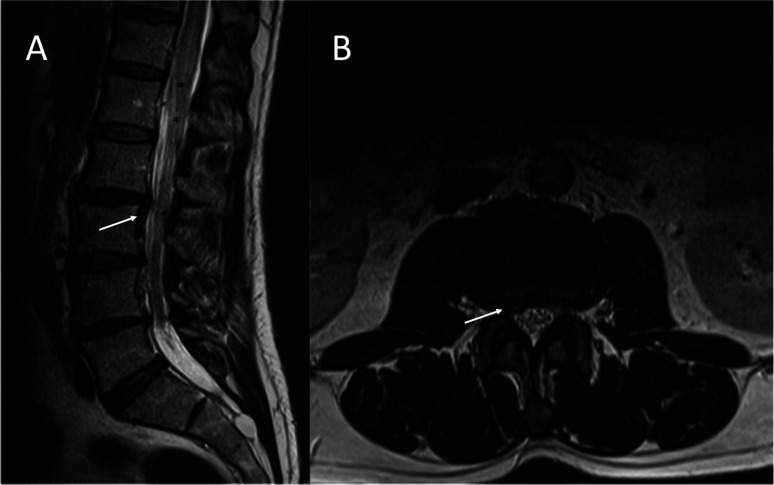
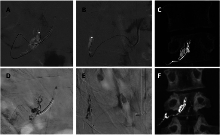
Spinal arteriovenous fistulas (SAVFs) are the most common type of vascular malformation of the spine in adult patients. They can lead to acute or progressive myelopathy due to venous congestion of the medullary veins. While most SAVFs are acquired, their pathophysiology remains unclear. The natural history of the disease and its clinical presentation are highly influenced by the location of the fistula and various factors may trigger sudden neurological decline. We present a case of a patient who developed a complete spinal cord injury after a lumbar nerve root block, likely due to an undiagnosed SAVF. The patient underwent endovascular embolization, resulting in a complete recovery of neurological function.

Keywords: Angiography; arteriovenous fistula; dural fistula; spine.


Conflict of interest statement

Declaration of conflicting interestsThe authors declared no potential conflicts of interest with respect to the research, authorship, and/or publication of this article.

Figures

Figure 1.
Figure 1.
Lumbosacral magnetic resonance imaging prior to lumbar puncture. A sagittal (A) and axial (B) T2-weighted were reported as revealing a right caudal migrated L2–L3 disc herniation (white arrows). In retrospect, this was an underdiagnosed spinal epidural arteriovenous fistula, and abnormal flow voids (black asterisks) went unnoticed.
Figure 2.
Figure 2.
Lumbosacral magnetic resonance imaging following lumbar puncture. A sagittal short tau inversion recovery (STIR) image shows intramedullary edema extending from T7 to the spinal cord cone (black asterisks from T8 to cord cone) and perimedullar vascular engorgement (white arrow).
Figure 3.
Figure 3.
Spinal catheter angiography. (A) and (B) Selective right L3 radicular artery angiogram in the AP and L views, respectively. A Simmons 5F catheter is located at the ostium of the artery (white arrow). A venous pouch (white circle) is observed, from which a bridging vein (black arrow) emerges, communicating with the ventral superficial medullary veins (black dotted box). (C) and (D) 3D reconstruction images in the AP and L views, respectively. Road map in the anteroposterior (E) and lateral views (F) showing two retrocorporeal arteries (white arrows) shunting the venous pouch (white circle). Augmented AP (G) and L (H) views displayed the fistulous points (white asterisks). 3D axial reconstruction image shows the arterial feeders joining the venous pouch at the lateral recess.
Figure 4.
Figure 4.
AP (A), L (B) views, and 3D reconstruction image (C) of the arterial feeders microcatheterization (white asterisks). Fluoroscopic AP (D), L (E), and 3D reconstruction images (F) show the glue cast in the venous pouch.
Figure 5.
Figure 5.
Anteroposterior (A) and lateral (B) views of the post-embolization control. Selective right L3 radicular artery angiogram demonstrated complete exclusion of the fistula (white circle).
Figure 6.
Figure 6.
Follow-up lumbosacral STIR MRI. At 4 months (A) intramedullary extended from T10 to the spinal cord cone (black asterisks). At 1-year (B) and 2-year follow-ups (C), MRI revealed complete remission of both the medullar edema and vascular engorgement.

References

    1. Alhendawy I, Homapour B, Chandra RV, et al. Acute paraplegia in patient with spinal dural arteriovenous fistula after lumbar puncture and steroid administration: A case report. Int J Surg Case Rep 2021; 81: 105797. - PMC - PubMed
    1. Petrin Z, Marino RJ, Oleson CV, et al. Paralysis after lumbar interlaminar epidural steroid injection in the absence of hematoma: A case of congestive myelopathy due to spinal dural arteriovenous Fistula and a review of the literature. Am J Phys Med Rehabil 2020; 99: e107–e110. - PubMed
    1. International Spine Intervention Society. In: Bogduk N. (ed) ISIS practice guidelines for spinal diagnostic and treatment procedures, 2nd ed. San Francisco: International Spine Intervention Society, 2013.
    1. Geibprasert S, Pereira V, Krings T, et al. Dural arteriovenous shunts: A new classification of craniospinal epidural venous anatomical bases and clinical correlations. Stroke 2008; 39: 2783–2794. - PubMed
    1. Mull M, Othman A, Dafotakis M, et al. Spinal epidural arteriovenous fistula with perimedullary venous reflux: Clinical and neuroradiologic features of an underestimated vascular disorder. AJNR Am J Neuroradiol 2018; 39: 2095–2102. - PMC - PubMed

LinkOut - more resources