. 2024 Aug 28;44(8):185.
doi: 10.1007/s10875-024-01788-5.

Hereditary C1q Deficiency is Associated with Type 1 Interferon-Pathway Activation and a High Risk of Central Nervous System Inflammation

Affiliations

Hereditary C1q Deficiency is Associated with Type 1 Interferon-Pathway Activation and a High Risk of Central Nervous System Inflammation

Clément Triaille et al. J Clin Immunol. .

Abstract

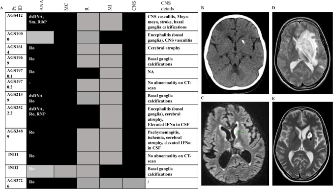
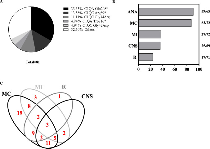
Hereditary C1q deficiency (C1QDef) is a rare monogenic disorder leading to defective complement pathway activation and systemic lupus erythematosus (SLE)-like manifestations. The link between impairment of the complement cascade and autoimmunity remains incompletely understood. Here, we assessed type 1 interferon pathway activation in patients with C1QDef. Twelve patients with genetically confirmed C1QDef were recruited through an international collaboration. Clinical, biological and radiological data were collected retrospectively. The expression of a standardized panel of interferon stimulated genes (ISGs) in peripheral blood was measured, and the level of interferon alpha (IFNα) protein in cerebrospinal fluid (CSF) determined using SIMOA technology. Central nervous system (encompassing basal ganglia calcification, encephalitis, vasculitis, chronic pachymeningitis), mucocutaneous and renal involvement were present, respectively, in 10, 11 and 2 of 12 patients, and severe infections recorded in 2/12 patients. Elevated ISG expression was observed in all patients tested (n = 10/10), and serum and CSF IFNα elevated in 2/2 patients. Three patients were treated with Janus-kinase inhibitors (JAKi), with variable outcome; one displaying an apparently favourable response in respect of cutaneous and neurological features, and two others experiencing persistent disease despite JAKi therapy. To our knowledge, we report the largest original series of genetically confirmed C1QDef yet described. Additionally, we present a review of all previously described genetically confirmed cases of C1QDef. Overall, individuals with C1QDef demonstrate many characteristics of recognized monogenic interferonopathies: particularly, cutaneous involvement (malar rash, acral vasculitic/papular rash, chilblains), SLE-like disease, basal ganglia calcification, increased expression of ISGs in peripheral blood, and elevated levels of CSF IFNα.

Keywords: C1Q deficiency; Complement; Janus-kinase inhibition; interferon; neuroinflammation; systemic lupus erythematosus.


Conflict of interest statement

The authors declare no competing interests.

Figures

Fig. 1
Fig. 1
Clinical Features of patients with C1QDef in our cohort. A For each patient, presence (dark) or absence (grey) of the following features is indicated: Anti-nuclear antibody (ANA- specific antibodies are written in the cell if present), mucocutaneous (MC), renal (R), major infection (MI) or central nervous system (CNS) involvement. Black and grey boxes indicate, respectively, the presence or absence of disease. Details of CNS involvement are given in the last column. CT-scan of patient AGS412 showing basal ganglia calcification. MRI of patient AGS2522.2 showing encephalitis with signal abnormalities in the basal ganglia and thalami (MRI sequence T2 FLAIR). MRI (T2W) of patient AGS1000 at relapse of CNS inflammation, showing diffuse enlargement of the left basal ganglia, caudate nucleus and thalamus, with mass effect. MRI (T2W) of patient AGS1000 two months after (D), showing significant reduction in the size of the lesion and perilesional oedema, and post biopsy changes
Fig. 2
Fig. 2
ISG expression in peripheral blood of C1QDef. A Expression of 6 ISGs in peripheral blood of n = 9 C1QDef patients compared to controls. ISG expression was determined either by qPCR (n = 8 patients), or NanoString (n = 1 patient). Age at sampling (years) and Interferon score are shown next to each patient ID. B Interferon score of n = 426 samples from canonical monogenic interferonopathies (results are grouped by mutant genotype), n = 79 controls and n = 9 C1QDef patients. Black: controls, red: elevated IS, blue: patient with IS in the range of controls. Whiskers show mean ± error of samples analysed using qPCR. Of note, ISG expression data of patient AGS3489 are not shown here (see Methods)
Fig. 3
Fig. 3
Clinical and radiological effects of JAKi in a patient with C1QDef. A-D Clinical picture of the face of patient AGS3489 and foot 2 weeks before (A-B) and 9 months after (C-D) initiation of treatment with baricitinib. The complete list of therapies at each time point is given in Supplementary Table 2. E–F Cerebral MRI (T1 sequence with contrasts) of patient AGS3489 3 months before initiation of baricitinib showing cerebral atrophy and pachymeningitis (arrows indicate meningeal thickening with enhancement). G-H Evolution of MRI after 6 months of baricitinib showing improvement in meningeal thickening and enhancement. Of note, a subdural hematoma appeared after lumbar puncture
Fig. 4
Fig. 4
Review of clinical features and genotypes of n = 77 published cases with genetically confirmed C1QDef. A Most frequent variants reported in C1QDef (73 homozygous and 4 compound heterozygous individuals). B Rate of ANA positivity (ANA), mucocutaneous manifestations (MC), major infections (MI), CNS (CNS) and renal involvement in C1QDef. The absolute numbers of cases with data available for each feature is shown on the right. C Venn diagram showing the co-occurrence of cardinal features of C1QDEF in n = 68 patients with data available for all cardinal features. Only 3 patients were diagnosed without any of these features

References

    1. Stegert M, Bock M, Trendelenburg M. Clinical presentation of human C1q deficiency: How much of a lupus? Mol Immunol. 2015;67(1):3–11. - PubMed
    1. van Schaarenburg RA, Schejbel L, Truedsson L, Topaloglu R, Al-Mayouf SM, Riordan A, et al. Marked variability in clinical presentation and outcome of patients with C1q immunodeficiency. J Autoimmun. 2015;62:39–44. - PubMed
    1. Coss SL, Zhou D, Chua GT, Aziz RA, Hoffman RP, Wu YL, et al. The complement system and human autoimmune diseases. J Autoimmun. 2023;137: 102979. - PMC - PubMed
    1. Santer DM, Hall BE, George TC, Tangsombatvisit S, Liu CL, Arkwright PD, et al. C1q deficiency leads to the defective suppression of IFN-alpha in response to nucleoprotein containing immune complexes. J Immunol. 2010;185(8):4738–49. - PMC - PubMed
    1. Meyer O, Hauptmann G, Tappeiner G, Ochs HD, Mascart-Lemone F. Genetic deficiency of C4, C2 or C1q and lupus syndromes. Association with anti-Ro (SS-A) antibodies. Clin Exp Immunol. 1985;62(3):678–84. - PMC - PubMed

Publication types

LinkOut - more resources