. 2024 Sep 11;146(36):24788-24799.
doi: 10.1021/jacs.3c12389. Epub 2024 Aug 28.

Targeted Protein Localization by Covalent 14-3-3 Recruitment

Affiliations

Targeted Protein Localization by Covalent 14-3-3 Recruitment

Qian Shao et al. J Am Chem Soc. .

Abstract

14-3-3 proteins have a unique ability to bind and sequester a multitude of diverse phosphorylated signaling proteins and transcription factors. Many previous studies have shown that interactions of 14-3-3 with specific phosphorylated substrate proteins can be enhanced through small-molecule natural products or fully synthetic molecular glue interactions. However, enhancing 14-3-3 interactions with both therapeutically intractable transcription factor substrates and potential neo-substrates to sequester and inhibit their function remains elusive. One of the 14-3-3 proteins, 14-3-3σ or SFN, has cysteine C38 at the substrate-binding interface, near the sites where previous 14-3-3 molecular glues have been found to bind. In this study, we screen a fully synthetic cysteine-reactive covalent ligand library to identify molecular glues that enhance the interaction of 14-3-3σ with not only druggable transcription factors such as estrogen receptor (ERα) but also challenging oncogenic transcription factors such as YAP and TAZ, which are part of the Hippo transducer pathway. We identify a hit EN171 that covalently targets both C38 and C96 on 14-3-3 to enhance 14-3-3 interactions with ERα, YAP, and TAZ, leading to impaired estrogen receptor and Hippo pathway transcriptional activity. We further demonstrate that EN171 could not only be used as a molecular glue to enhance native protein interactions but could also be used as a covalent 14-3-3 recruiter in heterobifunctional molecules to sequester nuclear neo-substrates such as BRD4 and BLC6 into the cytosol. Overall, our study reveals a covalent ligand that acts as a novel 14-3-3 molecular glue for challenging transcription factors such as YAP and TAZ and demonstrates that these glues can be potentially utilized in heterobifunctional molecules to sequester nuclear neo-substrates out of the nucleus and into the cytosol to enable targeted protein localization.


Conflict of interest statement

Competing Financial Interests Statement

DKN is a co-founder, shareholder, and scientific advisory board member for Frontier Medicines and Vicinitas Therapeutics. DKN is a member of the board of directors for Vicinitas Therapeutics. DKN is also on the scientific advisory board of The Mark Foundation for Cancer Research, Photys Therapeutics, Oerth Bio, Apertor Pharmaceuticals, and Deciphera Therapeutics. DKN is also an Investment Advisory Partner for a16z Bio, an Advisory Board member for Droia Ventures, and an iPartner for The Column Group.

Figures

Figure 1.
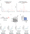
Figure 1.. Identification of 14-3-3 molecular glue stabilizers.
(a) Fluorescence polarization (FP) screening of a cysteine-reactive covalent ligand library (125 μM) alongside positive control Fusicoccin A (125 μM) against DMSO vehicle treatment of human 14-3-3σ protein (300 nM) and the phosphorylated ERα peptide (20 nM). The top hit, EN171, is shown in red. Individual compound and data for the screen can be found in Table S1. (b) Structure of EN171 with the cysteine-reactive acrylamide moiety highlighted in red. (c) EN171 showed an EC50 of 9 μM on 14-3-3σ with ERα, 45 μM with YAP, and 107 μM with TAZ. 14-3-3σ protein (300 nM) and the phosphorylated peptide (20 nM) were incubated with EN171 for 1 h. (d) Attenuation of EN171-mediated stabilization between 14-3-3σ and ERα, YAP, or TAZ with the 14-3-3σ inhibitor R18. 14-3-3σ protein (300 nM), the phosphorylated peptide (20 nM), and 14-3-3σ inhibitor R18 (100 μM) were incubated with EN171 for 4 h (ERα) or 1 h (TAZ and YAP). EC50 values for EN171-treatment conditions are also shown. EC50s could not be calculated for the R18 and EN171 treatment groups. Data shown in (c) and (d) are individual replicates (shown as staggered dots) from n=3 biologically independent replicates per group.
Figure 2.
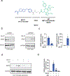
Figure 2.. Characterization of 14-3-3 Covalent Stabilizer EN171.
(a) Mass spectrometry analysis of EN171 modification on 14-3-3σ. Pure human protein 14-3-3σ with phosphorylated ERα peptide was incubated with EN171 (50 μM) for 30 min and tryptic digests from the protein were subsequently analyzed for the EN171 covalent adduct on a cysteine. Shown is the MS/MS data for EN171 modification on 14-3-3σ C38 and C96. (b) Structure of the alkyne-functionalized analog of EN171 (EN171-alkyne). (c) Gel-based ABPP showing that mutation of both C38 and C96 to serines completely ablated EN171-alkyne probe labeling. 14-3-3σ WT or mutant protein were incubated with EN171-alkyne (50 μM) for 1 hour. (d) Quantification of experiment in (c). (e, f) FP assay showing that mutation of either C38 or C96 to serines also significantly attenuated EN171-mediated stabilization of 14-3-3σ interaction with phosphorylated ERα (e) and TAZ (f) peptide substrates. 14-3-3σ WT or mutations (300 nM) and the phosphorylated peptide (20 nM) were incubated with EN171 for 4 hours. EC50 values for EN171-treatment conditions with WT 14-3-3 are also shown. EC50 values for EN171 treatment with mutant protein could not be derived across the data in (e, f) so they are not shown. Data shown in (c-f) are from n=3 biologically independent replicates per group. Data shown in (d,e,f) are individual replicate values and average ± sem for (d) and average values for (e,f) and significance is shown as *p<0.05 compared to WT 14-3-3 treatment groups.
Figure 3.
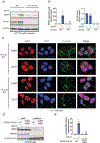
Figure 3.. Cellular Activity of EN171.
(a,b) Pulldown of endogenous 14-3-3σ in T47D cells using EN171-alkyne. T47D cells were treated with DMSO vehicle or EN171-alkyne probe (50 μM) for 1 h. An azide-functionalized biotin handle was appended onto probe-labeled proteins by CuAAC and these proteins were subsequently streptavidin-enriched. Resulting enriched proteins were analyzed by Western blotting for 14-3-3σ and loading control GAPDH and quantified in (b). (c) EN171-competed targets from EN171-alkyne pulldown proteomics. T47D cell lysate were spiked with 14-3-3σ pure protein, pre-treated with DMSO vehicle or EN171 (200 μM) for 1 h at 25 °C prior to EN171-alkyne probe labeling (40 μM) at 4 °C overnight. Probe-modified proteins were subjected to CuAAC with an azide-functionalized biotin and enriched by streptavidin after which eluates were analyzed by TMT-based quantitative proteomics. Shown in red are proteins that were significantly out-competed by EN171 by >2-fold with p<0.03. 14-3-3 is labeled with the larger red dot. Data are from n=3 biologically independent replicates per group. (d,e) Luciferase transcriptional reporter assays showing that EN171 dose-responsively impaired ERα and YAP/TAZ-mediated Hippo transcriptional activity in T47D and MCF7 breast cancer cells, respectively. Cells were treated with DMSO vehicle or EN171 for 24h. (f,g) Cellular ternary complex formation between 14-3-3σ and ERα with EN171 treatment. T47D cells expressing FLAG-14-3-3σ WT or C38/C96S protein were treated with DMSO vehicle or EN171 (100 μM) for 24h after which lysates were subjected to FLAG pulldown and enriched proteins were eluted and blotted for ERα, negative control GAPDH, and 14-3-3σ and both input and pulldown eluate protein levels were assessed by Western blotting and quantified in (g). (h,i) 14-3-3σ knockdown with shRNA in T47D and MCF7 cells assessed by Western blotting and quantified. (j,k) Attenuation of EN171-mediated inhibition of ERα and Hippo transcriptional luciferase reporter activity upon knockdown of 14-3-3σ. shControl and sh14-3-3σ T47D (j) and MCF7 (k) cells were treated with DMSO vehicle or EN171 (10 μM) for 24h. Data shown in (a,f,h,i) are representative of n=2–3 biologically independent replicates per group. Bar graphs in (d,e,f,g,h,i,j,k) show individual replicate values and average ± sem for (d,e,g,j,k) and average values for (h.i). Significance is expressed as *p<0.05 compared to vehicle-treated controls or vehicle-treated shControl cells in (b,d,e,g,h,i.j.k), and #p<0.05 compared to EN171 treated WT 14-3-3 protein in (g) and shControl cells in (j,k).
Figure 4.
Figure 4.. Heterobifunctional Recruiter for 14-3-3σ to Sequester Nuclear Neo-Substrate BRD4 in the Cytosol.
(a) Structure of QS-57 consisting of the covalent 14-3-3σ recruiter EN171 in blue linked via a C2 alkyl linker in grey to the BET family inhibitor JQ1 in green. (b,c) Sequestration of neo-substrate proteins BRD4 from the nucleus into the cytosol. T47D cells were treated with DMSO vehicle or QS-57 (25 μM) for 24 h. Nuclear and cytosolic fractions were separated and BRD4 and nuclear and cytosolic loading controls histone and GAPDH, respectively, were assessed by Western blotting (b) and quantified in (c). (d,e) Attenuation of BRD4 cytosolic sequestration by JQ1 pre-treatment. HCT116 cells were pre-treated with DMSO vehicle or JQ1 (100 μM) for 6 h prior to treatment of cells with DMSO vehicle or QS-57 for 24 h. Nuclear and cytosolic fractions were isolated and cytosolic BRD4 and loading control GAPDH were assessed by Western blotting and quantified in (e). Blots shown in (b,d) are representative of n=3 biologically independent replicates per group. Individual replicate values and average ± sem are shown in bar graphs from (c,e). Significance is expressed as *p<0.05 compared to vehicle-treated controls or vehicle-treated WT cells and #p<0.05 compared to QS-57-treated cells in (e).
Figure 5.
Figure 5.. Validating mechanism of BRD4 cytosolic sequestration by QS-57.
(a,b) Attenuation of BRD4 cytosolic sequestration by 14-3-3σ knockout (KO). Wild-type (WT) and 14-3-3σ KO HCT116 cells were treated with DMSO vehicle or QS-57 (100 μM) for 24h. Nuclear and cytosolic fractions were isolated and cytosolic BRD4, 14-3-3σ, and loading control GAPDH were assessed by Western blotting (a) and quantified in (b). (c) Imaging of nuclear and cytosolic BRD4 in cells. HCT116 14-3-3σ WT and KO cells were treated with DMSO vehicle or QS-57 (100 μM) for 3h, after which cells were fixed and imaged for BRD4 (in red), nuclear marker DAPI (in blue), cytosolic actin marker Phalloidin (in green) by microscopy. (d,e) Cellular ternary complex formation between 14-3-3σ and BRD4 with QS-57 treatment. HEK293 cells expressing FLAG-14-3-3σ WT or C38/C96S protein were treated with DMSO vehicle or QS-57 (100 μM) for 24h after which lysates were subjected to FLAG pulldown and enriched proteins were eluted and blotted for BRD4, negative control GAPDH, and 14-3-3 and both input and pulldown eluate protein levels were assessed by Western blotting and quantified in (e). Blots shown in (a,d) are representative of n=3 biologically independent replicates per group. Individual replicate values and average ± sem are shown in bar graphs from (b,e). Significance is expressed as *p<0.05 compared to vehicle-treated controls and #p<0.05 compared to QS-57-treated WT groups in (b,e).
Figure 6.
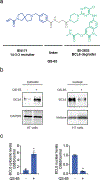
Figure 6.. Cytosolic sequestration of nuclear neo-substrate BCL6.
(a) Structure of QS-65 linking 14-3-3σ recruiter EN171 in blue via a C2 alkyl linker in grey to the BCL6 degrader BI-3802 in green. (b,c) Sequestration of BCL6 from the nucleus into the cytosol. HT cells were treated with DMSO vehicle or QS-65 (100 μM) for 15 h. Nuclear and cytosolic fractions were separated and BCL6 and nuclear and cytosolic loading controls histone and GAPDH, respectively, were assessed by Western blotting and quantified in (c). Blots shown in (b) are representative of n=3 biologically independent replicates per group. Individual replicate values and average ± sem are shown in bar graph in (c). Significance is expressed as *p<0.05 compared to vehicle-treated controls.

References

    1. Andrei SA et al. Stabilization of protein-protein interactions in drug discovery. Expert Opin. Drug Discov. 12, 925–940 (2017). - PubMed
    1. Ballone A, Centorrino F & Ottmann C 14-3-3: A Case Study in PPI Modulation. Mol. Basel Switz. 23, E1386 (2018). - PMC - PubMed
    1. Milroy L-G, Grossmann TN, Hennig S, Brunsveld L & Ottmann C Modulators of protein-protein interactions. Chem. Rev. 114, 4695–4748 (2014). - PubMed
    1. Ottmann C et al. A structural rationale for selective stabilization of anti-tumor interactions of 14-3-3 proteins by cotylenin A. J. Mol. Biol. 386, 913–919 (2009). - PubMed
    1. Ballone A, Centorrino F, Wolter M & Ottmann C Structural characterization of 14-3-3ζ in complex with the human Son of sevenless homolog 1 (SOS1). J. Struct. Biol. 202, 210–215 (2018). - PubMed