. 2024 Oct 1;35(10):ar134.
doi: 10.1091/mbc.E24-04-0179. Epub 2024 Aug 28.

The RhoGEF protein Plekhg5 self-associates via its PH domain to regulate apical cell constriction

Affiliations

The RhoGEF protein Plekhg5 self-associates via its PH domain to regulate apical cell constriction

Ivan K Popov et al. Mol Biol Cell. .

Abstract

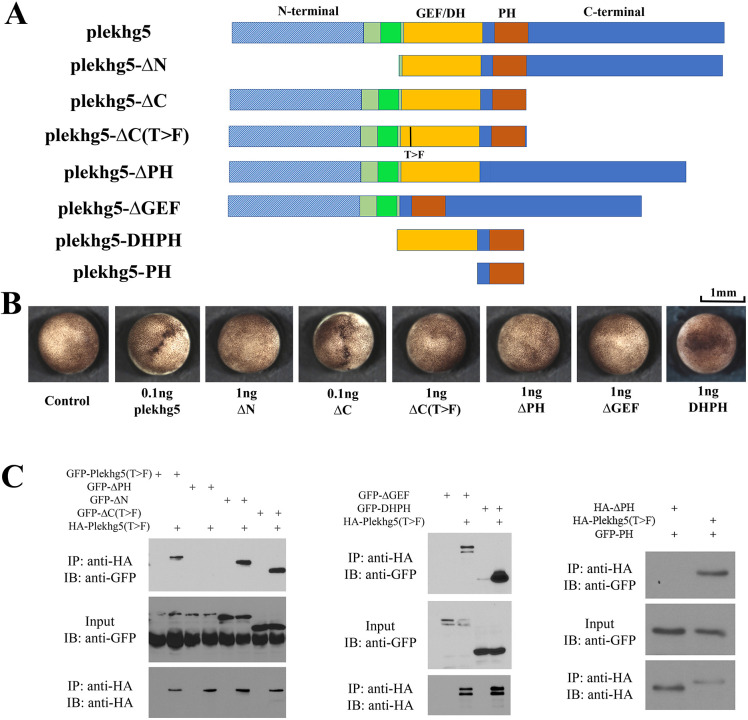
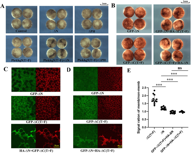
RhoGEFs are critical activators of Rho family small GTPases and regulate diverse biological processes, such as cell division and tissue morphogenesis. We reported previously that the RhoGEF gene plekhg5 controls apical constriction of bottle cells at the blastopore lip during Xenopus gastrulation, but the detailed mechanism of plekhg5 action is not understood in depth. In this study, we show that localization of Plekhg5 in the apical cortex depends on its N-terminal sequences and intact guanine nucleotide exchange activity, whereas the C-terminal sequences prevent ectopic localization of the protein to the basolateral compartment. We also reveal that Plekhg5 self-associates via its PH domain, and this interaction leads to functional rescue of two mutants that lack the N-terminal region and the guanine nucleotide exchange factor activity, respectively, in trans. A point mutation in the PH domain corresponding to a variant associated with human disease leads to loss of self-association and failure of the mutant to induce apical constriction. Taken together, our results suggest that PH-mediated self-association and N-terminal domain-mediated subcellular localization are both crucial for the function of Plekhg5 in inducing apical constriction.


Conflict of interest statement

Conflicts of interests: The authors declare no financial conflict of interest.

Figures

FIGURE 1:
FIGURE 1:
Both long and short isoforms of Plekhg5 induce apical constriction and can self-associate. (A) Schematic representation of Plekhg5 isoforms, T>F mutants, and the deletion mutants. The long isoform corresponds to the NCBI protein with the accession number XP_018083458, whereas the short isoform has the accession number of XP_018083456. (B) Induction of apical constriction by the wild-type (total 0.1 ng RNA), but not the T>F mutants (total 1 ng RNA), plekhg5 isoforms. The experiment was repeated three times, with the total number of embryos displaying apical constriction as follows: control, 0/28 embryos; 0.1 ng plekhg5, 28/28 embryos; 0.1 ng plekhg5-S, 28/28 embryos; 1 ng plekhg5(T>F), 0/39 embryos; and 1 ng plekhg5(T>F-S), 0/33 embryos. (C) Removal of the divergent N-terminal sequences in the isoforms (tN1 mutant) and including the first 44 amino acids in the overlapping region upstream of the GEF/DH domain (tN2 mutant) does not impair the ability of the mutants to induce apical constriction. The experiment was repeated three times, with the total number of embryos displaying apical constriction as follows: control, 0/42 embryo; 0.1 ng plekhg5, 23/23 embryos; 0.1 ng plekhg5(tN1), 44/44 embryos; and 0.1 ng plekhg5(tN2), 51/51 embryos. (D) Deletion of the 65 amino acids in the region common to long and short isoforms upstream of the GEF/DH domain leads to loss of induction of apical constriction by the mutant. The experiment was repeated three times, with the total number of embryos displaying apical constriction as follows: control, 0/38 embryos; 0.1 ng plekhg5, 26/26 embryos; and 1 ng plekhg5(Δ65), 0/45 embryos. (E) Both long and short isoforms of Plekhg5 can self-associate. HA- and GFP-tagged Plekhg5(T>F) or Plekhg5(T>F-S) were coexpressed at the RNA doses of 0.5 to 1 ng for each gene in early Xenopus embryos. co-IP studies were performed using embryonic lysate obtained at gastrula stages. co-IP signals were detected by Western blot analysis. The experiment was repeated three times. IP: immunoprecipitation; IB: immunoblotting.
FIGURE 2:
FIGURE 2:
The PH domain is critical for Plekhg5 self-association. (A) Schematic representation of the deletion mutants used in the co-IP assay. (B) The deletion mutants have different abilities to induce apical constriction. While removal of the C-terminal sequences (ΔC mutant) does not impair the ability of the mutant to induce apical constriction, deletion of the other domains leads to reduction (in the case of the DHPH mutant) or loss of induction of apical constriction by the mutants. The experiments were repeated three to five times, with the total number of embryos displaying apical constriction as follows: control, 0/44 embryos; 0.1 ng plekhg5, 61/61 embryos; 1 ng ΔN, 0/39 embryos; 0.1 ng ΔC, 61/61 embryos; 1 ng ΔC(T>F), 0/60 embryos; 1 ng ΔPH, 0/39 embryos; 1 ng ΔGEF, 0/51 embryos and 1 ng DHPH, 35/72 embryos with weak constriction. (C) Left panel: the co-IP studies indicate that deletion of the PH domain abolishes Plekhg5 self-association whereas deletion of the other domains does not affect association of the mutants with Plekhg5(T>F). Middle panel: deletion of the GEF/DH domain does not affect binding of the mutant to Plekhg5(T>F), and the isolated DHPH domain is sufficient for association of the mutant with Plekhg5(T>F). Right panel: the isolated PH domain is capable of binding to Plekhg5(T>F), but not the Plekhg5 mutant with the PH domain removed. The experiments were repeated three times.
FIGURE 3:
FIGURE 3:
Localization of Plekhg5 and its mutants. (A) GFP-tagged Plekhg5 and its mutants were coexpressed with membrane-tethered mCherry (mem-mCherry) in the animal cells of early embryos. Examination of protein localization at early gastrula stages indicates that wild-type Plekhg5 accumulates under the apical membrane, whereas the T>F mutant is moderately enriched at the cell junction. Deletion of the C-terminal region enhances junctional recruitment of the ΔC(T>F) mutant as well as its ectopic localization to the basolateral compartment of outer epithelial cells and cell-cell contacts of the inner cells. Deletion of the N-terminal sequence results in more diffuse distribution of the ΔN mutant. The experiments were repeated three times for en face view samples and twice for side view samples, with the total number of imaged embryos as follows: 0.1–0.2 ng GFP-Plekhg5, 14 en face view and six side view samples; 0.2–0.5 ng GFP-Plekhg5(T>F), 13 en face view and 12 side view samples; 0.5 ng GFP-ΔN, 13 en face view and 14 side view samples; and 0.5 ng GFP-ΔC(T>F), 13 en face view and 12 side view samples. Arrows indicate signals under the apical surface of plekhg5-expressing cells or enriched at the cell junctions of cells expressing plekhg5(T>F) or ΔC(T>F) in the en face view, whereas arrowheads point to ectopic signals in the basolateral compartment of outer epithelial cells and at the cell-cell contacts of inner cells in the side view. (B) Quantification of GFP signals at cell contacts versus in the nonjunctional areas in samples with en face views support the observation of junctional enrichment of the T>F mutants. A total of nine samples from three experiments were analyzed for each sample group. *** indicates p-value less than 0.001, and ns means nonsignificant with the p-value = 0.06 between the indicated groups.
FIGURE 4:
FIGURE 4:
Functional complementation of localization-impaired and GEF-defective mutants with coexpression. (A) Complementation of the Plekhg5(T>F) mutant with the localization defective ΔN, but not the ΔPH, mutant in inducing apical constriction. The experiment was repeated twice, with the total number of embryos displaying apical constriction as follows: control, 0/24 embryos; 1 ng ΔN, 0/24 embryos; 1 ng ΔPH, 0/24 embryos; 1 ng Plekhg5(T>F), 0/24 embryos; 0.5+0.5 ng Plekhg5(T>F)+ΔN, 24/24 embryos; and 0.5+0.5 ng Plekhg5(T>F)+ΔPH, 0/24 embryos. (B) The ΔN and the ΔC(T>F) mutants functionally complement to induce apical constriction. The experiment was repeated four times, with the total number of embryos displaying apical constriction as follows: control, 0/50 embryos; 1 ng GFP-ΔN, 0/66 embryos; 1 ng GFP-ΔC(T>F), 0/66 embryos; 0.5+0.5 ng GFP-ΔN+HA-ΔC(T>F), 62/62 embryos; and 0.5+0.5 ng GFP-ΔC(T>F)+HA-ΔN, 63/63 embryos. (C and D) Rescue of apical cortex localization of the GFP-tagged mutants when they are coexpressed with the other mutant that could restore junctional recruitment or a functional GEF domain via self-association. The experiments were repeated three (panel D) to four (panel C) times, with the total number of embryos imaged with the shown patterns as follows: 0.5 ng GFP-ΔN, 22/22 samples; 0.5 ng GFP-ΔC(T>F), 21/21 samples; 0.25+0.25 ng or 0.5+0.5 ng GFP-ΔN+HA-ΔC(T>F), 15/18 samples; and 0.25+0.25 ng or 0.5+0.5 ng GFP-ΔC(T>F)+HA-ΔN, 23/24 samples. (E) Quantification of GFP signal at the cell membrane and nonmembrane areas indicated a statistically significant difference between mutant alone and the rescued groups. *** indicates p-value less than 0.001, and ns means nonsignificant with the p-value = 0.24 between the indicated groups.
FIGURE 5:
FIGURE 5:
Computational modeling of Plekhg5 DH/PH domains as a dimer. (A) Homology modeling based on the structure of Arhgef11 reveals PH-mediated dimerization of the DH and PH domains of Plekhg5. (B) Two groups of amino acids (red and green) are identified to be potentially important for self-association at the DH/PH dimer interface. Mutagenesis of three hydrophobic residues into charged arginine is indicated in the sequence. (C) The 3R mutant cannot induce apical constriction. The experiment was repeated three times, with the total number of embryos displaying apical constriction as follows: control, 0/24 embryos; 0.1 ng plekhg5, 32/32 embryos; 1 ng plekhg5(3R), 0/40 embryos. (D) When expressed at the similar protein level to Plekhg5(T>F), with RNA doses of 0.5 ng HA-plekhg5(T>F) and 2 ng HA-plekhg5(3R), the 3R mutant fails to self-associate in the co-IP assay. The experiment was repeated three times.
FIGURE 6:
FIGURE 6:
The PH mutants corresponding to those identified in human PLEKHG5 disease-associated variants have different abilities to induce apical constriction or self-associate. (A) Sequence alignment between human and Xenopus proteins demonstrates conservation of the residues found in disease-associated human variants in the PH domain. These residues are found in the beta-sandwiches or the loop regions in the predicted structure of the PH domain based on the AlphaFold model. (B) Distinct abilities of the Xenopus mutants corresponding to the human variants in inducing apical constriction. While the T609M and the T663M mutants retained their ability to induce apical constriction, the P630H and F647S mutants showed severe reduction in inducing apical constriction. The experiments were repeated four times, with the total number of embryos displaying apical constriction as follows: control, 0/78 embryos; 0.1 ng HA-plekhg5, 50/50 embryos; 0.1 ng HA-T609M, 75/77 embryos; 1 ng HA-P630H, 5/73 embryos; 1 ng HA-F647S, 0/72 embryos; and 0.1 ng HA-T663M, 88/88 embryos. (C) The co-IP assay reveals that the P630H mutant is less stable than the wild-type protein and the F647S mutant fails to associate with the GFP-tagged DHPH domain of Plekhg5. The RNA doses used were: 1 ng GFP-DHPH; 2 ng HA-P630H; and 0.5 ng all the rest of the HA-tagged Plekhg5 mutants. The experiment was repeated three times. (D) Unlike the wild-type PH domain of Plekhg5, the isolated PH domains of P630H and F647S mutants cannot bind to Plekhg5. The experiment was repeated twice.

References

    1. Aigouy B, Farhadifar R, Staple DB, Sagner A, Roper JC, Julicher F, Eaton S (2010). Cell flow reorients the axis of planar polarity in the wing epithelium of Drosophila. Cell 142, 773–786. - PubMed
    1. Azevedo D, Antunes M, Prag S, Ma X, Hacker U, Brodland GW, Hutson MS, Solon J, Jacinto A (2011). DRhoGEF2 regulates cellular tension and cell pulsations in the Amnioserosa during Drosophila dorsal closure. PLoS One 6, e23964. - PMC - PubMed
    1. Azzedine H, Zavadakova P, Plante-Bordeneuve V, Vaz Pato M, Pinto N, Bartesaghi L, Zenker J, Poirot O, Bernard-Marissal N, Arnaud Gouttenoire E, et al. (2013). PLEKHG5 deficiency leads to an intermediate form of autosomal-recessive Charcot-Marie-Tooth disease. Hum Mol Genet 22, 4224–4232. - PMC - PubMed
    1. Baldwin AT, Popov IK, Keller R, Wallingford JB, Chang C (2022). The RhoGEF protein Plekhg5 regulates medioapical actomyosin dynamics of apical constriction during Xenopus gastrulation. bioRxiv. - PMC - PubMed
    1. Banani SF, Lee HO, Hyman AA, Rosen MK (2017). Biomolecular condensates: Organizers of cellular biochemistry. Nat Rev Mol Cell Biol 18, 285–298. - PMC - PubMed

Substances

LinkOut - more resources