. 2024 Oct 7;21(10):4970-4982.
doi: 10.1021/acs.molpharmaceut.4c00299. Epub 2024 Aug 28.

Development and Characterization of a Peptide-Bisphosphonate Nanoparticle for the Treatment of Breast Cancer

Affiliations

Development and Characterization of a Peptide-Bisphosphonate Nanoparticle for the Treatment of Breast Cancer

Kimberley Glass et al. Mol Pharm. .

Abstract

In women, breast cancer (BC) is the most common cancer, and despite advancements in diagnosis and treatment, 20-30% of early stage BC patients develop metastatic disease. Metastatic BC is deemed an incurable disease, which accounts for 90% of BC related deaths, with only 26% of metastatic patients reaching a 5 year survival rate. Therefore, there is an unmet need for the prevention or treatment of metastasis in early stage breast cancer patients. Bisphosphonates (BPs) are potent inhibitors of bone resorption and are extensively used for the prevention of osteoporosis and other skeletal disorders, as well as for the treatment of secondary bone cancer in BC patients. Furthermore, the direct anticancer activity of BPs has been established in primary tumor models. However, these studies were limited by the need for dosages far above the clinical range to overcome BPs' high affinity for bones and poor accumulation in the tumor itself, which leads to toxicity, including osteonecrosis of the jaw. To decrease BP dosage, increase bioavailability, and direct anticancer activity, we used the RALA (R-) peptide delivery system to form highly stable NPs with the nitrogen containing BP, risedronate (R-RIS). In vitro studies showed that, in comparison to RIS, R-RIS nanoparticles increased cytotoxicity and reduced metastatic features such as proliferation, migration, invasion, and adhesion of metastatic BC cells to bones. Furthermore, in an in vivo model, R-RIS had increased tumor accumulation while still maintaining similar bone accumulation to RIS alone. This increase in tumor accumulation corresponded with decreased tumor volume and lungs metastasis. R-RIS has great potential to be used in combination with standard of care chemotherapy for the treatment of primary BC and its metastasis while still having its bone resorption inhibiting properties.

Keywords: RALA; bisphosphates; breast cancer; nanomedicine; risedronate.


Conflict of interest statement

The authors declare no competing financial interest.

Figures

Figure 1
Figure 1
R-RIS Nanoparticle formation and characterization (A) 1 nmol of RIS was complexed with increasing nmolar ratios of RALA peptide (0.2–2) and the hydrodynamic size, mean count rate, zeta potential and PDI were evaluated using a Zetasizer. A fixed molar ratio of 1:1 RALA/RIS (60 μM) was used for further characterization studies. (B,i) Zeta potential and hydrodynamic size, mean count rate and polydispersity index (PDI) of 1:1 ratio particles was measured over 12 months. (C) Size and PDI was measured at various temperatures. (D) Representative TEM image of 1:1 ratio particles. (E) Fluorescent (red) AF647-RIS was used to evaluate complexation and cellular uptake. Particles were formulated with 0.1 μg pf AF647-RIS at increasing nmolar ratios. Complexation was calculated using AF647-RIS only as a control with 100% fluorescence and 0% complexation. (F) MDA-MB-231 cells were either untreated or treated with either 0.1 μg uncomplexed AF647-RIS or RALA/AF647-RIS nanoparticle for 2 h. Slides were fixed, stained with phalloidin (green), and mounted with DAPI (blue) mounting medium. Slides were imaged using confocal microscopy. Arrow shows internalization of AF647-RIS (red). Scale bar = 49.1 μm. All results are displayed ± SEM, n = 3.
Figure 2
Figure 2
R-RIS in vitro cytotoxicity. (A) MDA-MB-231, MCF7 and 4T1 BC cell lines were treated with 1:1 nmolar ratio RIS/R-RIS particles for 6 h before replacement with complete media. Viability was assessed 72 h later via alamarBlue. Mann–Whitney unpaired two tailed test, sum of square F test was applied. (B) Cells were treated with 20 μM RIS/R-RIS for 6 h and then seeded in 6 well plates and left for 7 (4T1) or 14 days (MDA-MB-231, MCF-7) and stained with crystal violet. The SF was determined. A Kruskal–Wallis nonparametric test was applied. (C) MDA-MB-231 cells were seeded at 2 × 10³ cells/well in a 96 well round-bottom plate pre-coated with 6 μL of 120 mg/mL Poly-Hema. Matrigel was added 24 h later in a final concentration of 2.5%. Spheroids were treated with 20–180 μM and left for 10 days. Spheroid viability was measured via the Alamar Assay. A Mann–Whitney two tailed test was applied. (D) Spheroids were treated on day 4 with 160 μM RIS/R-RIS and the volume was measured over 10 days. A Kruskal–Wallis nonparametric test was applied. (E) MDA-MB-231 cells were treated with a range of RIS and R-RIS concentrations for 6 h, and protein was collected 48 h later. Protein was probed for caspase-3, cleaved caspase-3, and γ-tubulin as a loading control. (F) Cells were treated for 6 h and protein was collected. Protein was probed for pAkt, total Akt and GAPDH as a loading control. (G) Densitometry measurements were performed on expression of pAkt relative to control and normalized to total Akt. All results are displayed as ± SEM, n = 3. A Kruskal–Wallis nonparametric test was applied.
Figure 3
Figure 3
R-RIS ability to inhibit metastasis. (A) MDA-MB-231, MCF-5 and 4T1 cells were treated in a 24-well plate with 60 μM RIS/R-RIS for 6 h. A scratch was made using a p1000. Photos were taken at 24 and 48 h for MDA-MB-231 and MCF-7 cells and 6 and 24 h for 4T1 cells. The percent closure was measured. (B) MDA-MB-231 cells were treated for 6 h with 20–100 μM RIS/R-RIS and protein collected 48 h later. E-cadherin, Zeb2, N-cadherin, MMP2 and Vimentin were probed with γ-tubulin used as a loading control. (C) MDA-MB-231 cells were treated for 6 h with 20 μM RIS/R-RIS, spun down, and resuspended in serum free medium to ensure no FBS was present. 2 × 10⁵ cells were seeded in a Transwell chamber with 150 μL present in each well. Wells were stained at 24 and 48 h, and cell invasion was measured. (D) MG63 cells were seeded at 3 × 10⁵ cells/well in a 24-well plate. EGFP-transfected MDA-MB-231 cells were treated with 20 μM RIS/R-RIS for 6 h and seeded at 2 × 10⁵ cells/well on top of the MG63 monolayer. Cells were left for 1 h to adhere. Cells were washed, and fluorescence was read between 5 and 60 min to calculate the percentage of cell adherence. All results are displayed ± SEM, n = 3. A Kruskal–Wallis nonparametric test was applied in (A,B) and (D).
Figure 4
Figure 4
R-RIS accumulation in organs. AF647-RIS was used to assess accumulation in mouse tumor and bone. BALB/c SCID mice were implanted subcutaneously with 5 × 10⁶ MDA-MB-231 cells and grown to 150 mm3. Mice were treated intravenously with 10 μg of AF6476-RIS or RALA/AF647-RIS. Tumours and femurs were harvested after 1 h. (A) Fluorescent images of tumors and femurs. The relative mean drug fluorescence intensity of (B) tumors and (C) bone normalized to the weight of tumors and bone in untreated mice. (D) Organs. Results are displayed as ± SEM, n = 3. A Kruskal–Wallis nonparametric test was applied.
Figure 5
Figure 5
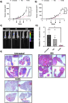
R-RIS in vivo cytotoxicity. (A) BALB/c SCID mice were implanted subcutaneously with 5 × 10⁶ MDA-MB-231 cells and grown to 150 mm3. Mice (n = 6 per group) were treated weekly intravenously with 10 μg AF6476-RIS or RALA/AF647-RIS. Tumour volume was measured. Arrows indicate when treatment occurred (week 2, 3, and 4). (B) BALB/c mice were implanted with 5 × 10⁴ 4T1 cells in the 4th mammary fat pad and tumors grew to 150 mm3 before treatment. Mice (n = 8 per group) were treated with 10 μg RIS or R-RIS intravenously weekly. Tumour volume was measured. Arrows indicate when mice were treated (week 2, 3, and 4). (C) Representative luminescent images showing 4T1 tumor and metastasis to the lungs. (D) Lungs were harvested and incubated in the presence of 6-thioguanine. After 7 days, resistant 4T1 cells were stained with crystal violet (Figure S8) and absorbance was measured 4 lungs per group were analyzed. (E) H&E staining was performed on 4T1 tumor bearing untreated, RIS treated, and R-RIS treated mouse lungs in the Pathology and Diagnostics lab in the Royal Veterinary Collage by Ms. Sue Rodway. Samples were imaged on a brightfield microscope at 60× magnification. Tumour lesions are highlighted in red boxes. Results are displayed as mean ± SEM. A Kruskal–Wallis nonparametric tests were applied.

References

    1. Early Breast Cancer (Preventing Recurrance and Improving Survival): Adjuvant Bisphosphonates [ES15]; National Institute for Health and Care Excellence, 2017.
    1. Redig A. J.; McAllister S. S. Breast cancer as a systemic disease: a view of metastasis. J. Intern. Med. 2013, 274 (2), 113–126. 10.1111/joim.12084. - DOI - PMC - PubMed
    1. Kuchuk I.; Hutton B.; Moretto P.; Ng T.; Addison C. L.; Clemons M. Incidence, consequences and treatment of bone metastases in breast cancer patients-Experience from a single cancer centre. J. Bone Oncol 2013, 2 (4), 137–144. 10.1016/j.jbo.2013.09.001. - DOI - PMC - PubMed
    1. Lorusso G.; Rüegg C. New insights into the mechanisms of organ-specific breast cancer metastasis. Semin. Cancer Biol. 2012, 22, 226–233. 10.1016/j.semcancer.2012.03.007. - DOI - PubMed
    1. Goldstein J. L.; Brown M. S. Regulation of the mevalonate pathway. Nature 1990, 343 (6257), 425–430. 10.1038/343425a0. - DOI - PubMed