. 2024 Oct 1;327(4):E533-E543.
doi: 10.1152/ajpendo.00246.2024. Epub 2024 Aug 28.

Enhanced ECCD36 signaling promotes skeletal muscle insulin resistance in female mice

Affiliations

Enhanced ECCD36 signaling promotes skeletal muscle insulin resistance in female mice

Austin Dada et al. Am J Physiol Endocrinol Metab. .

Abstract

Consumption of a Western diet (WD) increases CD36 expression in vascular, hepatic, and skeletal muscle tissues promoting lipid metabolic disorders and insulin resistance. We further examined the role of endothelial cell-specific CD36 (ECCD36) signaling in contributing to skeletal muscle lipid metabolic disorders, insulin resistance, and their underlying molecular mechanisms. Female ECCD36 wild-type (ECCD36+/+) and knock-out (ECCD36-/-) mice, aged 6 wk, were provided with either a WD or a standard chow diet for a duration of 16 wk. ECCD36+/+ WD mice were characterized by elevated fasting plasma glucose and insulin levels, increased homeostatic model assessment for insulin resistance, and glucose intolerance that was blunted in ECCD36-/- mice. Improved insulin sensitivity in ECCD36-/- mice was characterized by increased phosphoinositide 3-kinases/protein kinase B signaling that further augmented glucose transporter type 4 expression and glucose uptake. Meanwhile, 16 wk of WD feeding also increased skeletal muscle free fatty acid (FFA) and lipid accumulation, without any observed changes in plasma FFA levels. These lipid metabolic disorders were blunted in ECCD36-/- mice. Moreover, ECCD36 also mediated in vitro palmitic acid-induced lipid accumulation in cultured ECs, subsequently leading to the release of FFAs into the culture media. Furthermore, consumption of a WD increased FFA oxidation, mitochondrial dysfunction, impaired mitochondrial respiratory, skeletal muscle fiber type transition, and fibrosis. These WD-induced abnormalities were blunted in ECCD36-/- mice. These findings demonstrate that endothelial-specific ECCD36 signaling participates in skeletal muscle FFA uptake, ectopic lipid accumulation, mitochondrial dysfunction, insulin resistance, and associated skeletal muscle dysfunction in diet-induced obesity.NEW & NOTEWORTHY ECCD36 exerts "extra endothelial cell" actions in skeletal muscle insulin resistance. ECCD36 is a major mediator of Western diet-induced lipid metabolic disorders and insulin resistance in skeletal muscle. Mitochondrial dysfunction is associated with diet-induced CD36 activation and related skeletal muscle insulin resistance.

Keywords: CD36; endothelial cells; insulin resistance; obesity; skeletal muscle.


Conflict of interest statement

No conflicts of interest, financial or otherwise, are declared by the authors.

Figures

None
Graphical abstract
Figure 1.
Figure 1.
ECCD36 deletion prevents insulin resistance induced by 16 wk of WD feeding. Fasting plasma glucose (A) and insulin levels (B) were measured in mg/dL and ng/mL, respectively. n = 7–10. C: HOMA-IR was calculated based on fasting plasma glucose and insulin values. n = 7–10. D: in vivo glucose intolerance was determined by IPGTT. n = 7 or 8. ECCD36−/− mice prevented WD-induced reduction in skeletal muscle PI3K/Akt expression (E) with quantitative analysis (F and G). n = 6. †P < 0.05 vs. ECCD36+/+ CD. ‡P < 0.05 vs. ECCD36+/+ WD groups. CD, chow diet; HOMA-IR, homeostatic model assessment for insulin resistance; IPGTT, intraperitoneal glucose tolerance test; WD, Western diet.
Figure 2.
Figure 2.
ECCD36 participates in regulating GLUT4 expression. Protein expression of GLUT1, GLUT3, and GLUT4 was examined by immunoblotting (A) with quantitative analysis (B) (n = 6). C: representative immunohistochemistry images of GLUT4 expression with quantitative analysis in membrane GLUT4 (D). Scales bars = 50 µm. n = 5–7. †P < 0.05 vs. ECCD36+/+ CD. ‡P < 0.05 vs. ECCD36+/+ WD groups. CD, chow diet; WD, Western diet.
Figure 3.
Figure 3.
ECCD36 deletion attenuates skeletal muscle lipid metabolic disorders in diet-induced obesity. Sixteen weeks of WD increased plasma NEFA (A) (n = 10), ex vivo skeletal muscle FFA levels (B) (n = 5), and lipid accumulation determined by Oil red O staining (C and D) (n = 6 or 7) in ECCD36+/+ mice. Increased skeletal muscle FFA levels and lipid accumulation were blunted in WD-fed ECCD36−/− mice (B–D). Scale bars = 50 µm. E: in the coculture system with 0.1 mM PA-treated ECs, CD36-siRNA in ECs inhibited lipid-rich ECs (treated by PA)-induced increase in FFA levels in the bottom culture media. n = 5. †P < 0.05 vs. ECCD36+/+ CD or Control-siRNA. ‡P < 0.05 vs. ECCD36+/+ WD groups or PA+Control-siRNA. CD, chow diet; ECs, endothelial cells; FFA, free fatty acid; NEFA, nonesterified fatty acid; PA, palmitic acid; WD, Western diet.
Figure 4.
Figure 4.
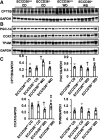
ECCD36 deletion inhibits FFA oxidation and prevents mitochondrial dysfunction. A: WD induced an increase in CPT1B that was blunted in ECCD36−/− mice. B: expression of mitochondrial encoded genes PGC-1α, COX1, and TFAM with quantitative analysis (C). n = 6. †P < 0.05 vs. ECCD36+/+ CD. ‡P < 0.05 vs. ECCD36+/+ WD groups. CD, chow diet; FFA, free fatty acid; WD, Western diet.
Figure 5.
Figure 5.
ECCD36 deletion inhibits impaired mitochondrial respiratory and oxygen consumption. A: ECCD36 signaling mediated the WD-induced reduction in ex vivo skeletal muscle protein expression of the respiratory complexes of the electron transport chain. Quantitative analysis is provided in B–F. G: ECCD36 mediated WD-induced reduction in ex vivo skeletal muscle mitochondrial oxygen consumption. n = 5 or 6. †P < 0.05 vs. ECCD36+/+ CD. ‡P < 0.05 vs. ECCD36+/+ WD groups. CD, chow diet; WD, Western diet.
Figure 6.
Figure 6.
ECCD36 deletion inhibits skeletal muscle fiber type transition, oxidative stress, and fibrosis. A: ECCD36 mediated WD-induced slow-to-fast muscle transitions in slow-twitch soleus muscle with quantitative analysis shown in B. n = 6. Representative images of immunostaining for skeletal muscle with 3 nitrotyrosine (C) and picrosirius (D) with quantitative analysis. Scales bars = 50 µm. n = 6 or 7. †P < 0.05 vs. ECCD36+/+ CD. ‡P < 0.05 vs. ECCD36+/+ WD groups. CD, chow diet; WD, Western diet.

References

    1. Bergman BC, Goodpaster BH. Exercise and muscle lipid content, composition, and localization: influence on muscle insulin sensitivity. Diabetes 69: 848–858, 2020. doi: 10.2337/dbi18-0042. - DOI - PubMed
    1. Jia D, Zhang J, Liu X, Andersen JP, Tian Z, Nie J, Shi Y. Insulin resistance in skeletal muscle selectively protects the heart in response to metabolic stress. Diabetes 70: 2333–2343, 2021. doi: 10.2337/db20-1212. - DOI - PMC - PubMed
    1. Reynolds LJ, Credeur DP, Manrique C, Padilla J, Fadel PJ, Thyfault JP. Obesity, type 2 diabetes, and impaired insulin-stimulated blood flow: role of skeletal muscle NO synthase and endothelin-1. J Appl Physiol (1985) 122: 38–47, 2017. doi: 10.1152/japplphysiol.00286.2016. - DOI - PMC - PubMed
    1. Friedman JE, Ishizuka T, Shao J, Huston L, Highman T, Catalano P. Impaired glucose transport and insulin receptor tyrosine phosphorylation in skeletal muscle from obese women with gestational diabetes. Diabetes 48: 1807–1814, 1999. doi: 10.2337/diabetes.48.9.1807. - DOI - PubMed
    1. Guglielmi V, Maresca L, D'Adamo M, Di Roma M, Lanzillo C, Federici M, Lauro D, Preziosi P, Bellia A, Sbraccia P. Age-related different relationships between ectopic adipose tissues and measures of central obesity in sedentary subjects. PLoS One 9: e103381, 2014. doi: 10.1371/journal.pone.0103381. - DOI - PMC - PubMed

LinkOut - more resources