

Developing a low-cost, open-source, locally manufactured workstation and computational pipeline for automated histopathology evaluation using deep learning
- PMID: 39197222
- PMCID: PMC11399610
- DOI: 10.1016/j.ebiom.2024.105276
Developing a low-cost, open-source, locally manufactured workstation and computational pipeline for automated histopathology evaluation using deep learning
Abstract
Background: Deployment and access to state-of-the-art precision medicine technologies remains a fundamental challenge in providing equitable global cancer care in low-resource settings. The expansion of digital pathology in recent years and its potential interface with diagnostic artificial intelligence algorithms provides an opportunity to democratize access to personalized medicine. Current digital pathology workstations, however, cost thousands to hundreds of thousands of dollars. As cancer incidence rises in many low- and middle-income countries, the validation and implementation of low-cost automated diagnostic tools will be crucial to helping healthcare providers manage the growing burden of cancer.
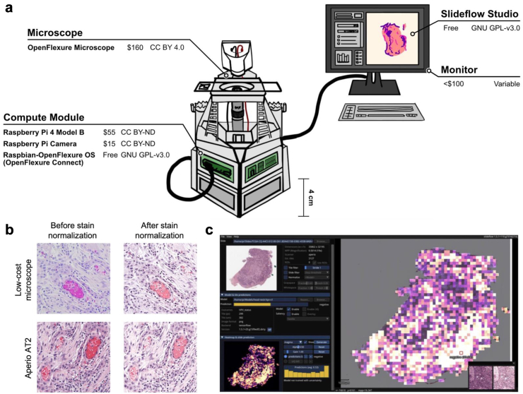
Methods: Here we describe a low-cost ($230) workstation for digital slide capture and computational analysis composed of open-source components. We analyze the predictive performance of deep learning models when they are used to evaluate pathology images captured using this open-source workstation versus images captured using common, significantly more expensive hardware. Validation studies assessed model performance on three distinct datasets and predictive models: head and neck squamous cell carcinoma (HPV positive versus HPV negative), lung cancer (adenocarcinoma versus squamous cell carcinoma), and breast cancer (invasive ductal carcinoma versus invasive lobular carcinoma).
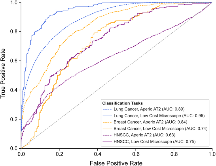
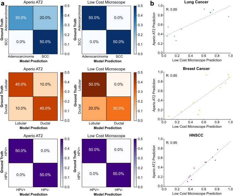
Findings: When compared to traditional pathology image capture methods, low-cost digital slide capture and analysis with the open-source workstation, including the low-cost microscope device, was associated with model performance of comparable accuracy for breast, lung, and HNSCC classification. At the patient level of analysis, AUROC was 0.84 for HNSCC HPV status prediction, 1.0 for lung cancer subtype prediction, and 0.80 for breast cancer classification.
Interpretation: Our ability to maintain model performance despite decreased image quality and low-power computational hardware demonstrates that it is feasible to massively reduce costs associated with deploying deep learning models for digital pathology applications. Improving access to cutting-edge diagnostic tools may provide an avenue for reducing disparities in cancer care between high- and low-income regions.
Funding: Funding for this project including personnel support was provided via grants from NIH/NCIR25-CA240134, NIH/NCIU01-CA243075, NIH/NIDCRR56-DE030958, NIH/NCIR01-CA276652, NIH/NCIK08-CA283261, NIH/NCI-SOAR25CA240134, SU2C (Stand Up to Cancer) Fanconi Anemia Research Fund - Farrah Fawcett Foundation Head and Neck Cancer Research Team Grant, and the European UnionHorizon Program (I3LUNG).
Keywords: Cancer diagnostics; Digital pathology; Global health; Low-cost microscope; Machine learning; Open-source; Precision oncology.
Copyright © 2024. Published by Elsevier B.V.
Conflict of interest statement
Declaration of interests A.T.P. reports no competing interests for this work, and reports personal fees from Prelude Therapeutics Advisory Board, Elevar Advisory Board, AbbVie consulting, Ayala Advisory Board, and stock options ownership in Privo Therapeutics, all outside of submitted work. J.M.D. is Founder/CEO of Slideflow Labs Inc, a digital pathology startup company founded in April 2024; he reports no financial interests related to the contents of this manuscript. S.R. is CSO of Slideflow Labs, owns stock/stock options in Slideflow Labs, and reports no competing interests for this work. F.M.H. reports receiving grants from the NIH/NCI, the Cancer Research Foundation, and the Department of Defense Breast Cancer Research Program and has no competing interests for this work. J.N.K. reports no competing interests for this work. He receives consulting fees from Owkin, DoMore Diagnostics, Panakeia, Scailyte, and Histofy, honoraria from AstraZeneca, Bayer, Eisai, MSD, BMS, Roche, Pfizer, and Fresenius, and reports owning stock/stock options in StratifAI GmbH. M.G. reports no competing interests for this work, and reports personal financial support from AstraZeneca, Abion, Merck Sharp & Dohme International GmbH, Bayer, Bristol-Myers Squibb, Boehringer Ingelheim Italia S.p.A, Celgene, Eli Lilly, Incyte, Novartis, Pfizer, Roche, Takeda, Seattle Genetics, Mirati, Daiichi Sankyo, Regeneron, Merck, Blueprint, Janssen, Sanofi, AbbVie, BeiGenius, Oncohost, and Medscape, Gilead, and Io Biotech.
Figures
References
-
- Fidler M.M., Bray F., Soerjomataram I. The global cancer burden and human development: a review. Scand J Public Health. 2018;46(1):27–36. - PubMed
-
- García-Rojo M. International clinical guidelines for the adoption of digital pathology: a review of technical aspects. Pathobiology. 2016;83(2–3):99–109. - PubMed
MeSH terms
LinkOut - more resources
Full Text Sources
Research Materials
