. 2024 Oct 3;187(20):5530-5539.e8.
doi: 10.1016/j.cell.2024.07.057. Epub 2024 Aug 27.

Animal and bacterial viruses share conserved mechanisms of immune evasion

Affiliations

Animal and bacterial viruses share conserved mechanisms of immune evasion

Samuel J Hobbs et al. Cell. .

Abstract

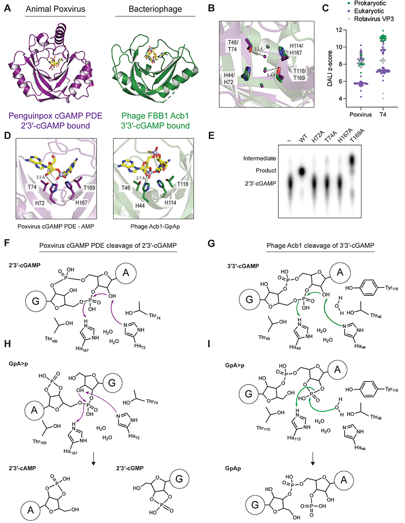
Animal and bacterial cells sense and defend against viral infections using evolutionarily conserved antiviral signaling pathways. Here, we show that viruses overcome host signaling using mechanisms of immune evasion that are directly shared across the eukaryotic and prokaryotic kingdoms of life. Structures of animal poxvirus proteins that inhibit host cGAS-STING signaling demonstrate architectural and catalytic active-site homology shared with bacteriophage Acb1 proteins, which inactivate CBASS anti-phage defense. In bacteria, phage Acb1 proteins are viral enzymes that degrade host cyclic nucleotide immune signals. Structural comparisons of poxvirus protein-2'3'-cGAMP and phage Acb1-3'3'-cGAMP complexes reveal a universal mechanism of host nucleotide immune signal degradation and explain kingdom-specific additions that enable viral adaptation. Chimeric bacteriophages confirm that animal poxvirus proteins are sufficient to evade immune signaling in bacteria. Our findings identify a mechanism of immune evasion conserved between animal and bacterial viruses and define shared rules that explain host-virus interactions across multiple kingdoms of life.

Keywords: CBASS; antiviral immunity; cGAS; cGLR; cyclic nucleotide; immune evasion; poxvirus.


Conflict of interest statement

Declaration of interests J.A.D. is a co-founder of Caribou Biosciences, Editas Medicine, Scribe Therapeutics, Intellia Therapeutics, and Mammoth Biosciences. J.A.D. is a scientific advisory board member of Vertex, Caribou Biosciences, Intellia Therapeutics, Scribe Therapeutics, Mammoth Biosciences, Algen Biotechnologies, Felix Biosciences, The Column Group, and Inari Agriculture. J.A.D. is Chief Science Advisor to Sixth Street, a Director at Johnson & Johnson, and Altos and Tempus and has research projects sponsored by Apple Tree Partners and Roche.

Figures

Figure 1.
Figure 1.. Animal poxvirus cGAMP PDE proteins are structural and functional homologs of bacteriophage Acb1 immune evasion enzymes
(A) Overview of the crystal structure of penguinpox cGAMP PDE in complex with 2’3’-cGAMP degradation products 3’-AMP and guanine (left). Overview of the crystal structure of bacteriophage FBB1 Acb1 in complex with the 3’3’-cGAMP degradation product GpAp (right). (B) Detailed view of the aligned catalytic residues of either animal poxvirus cGAMP PDE (purple) or phage Acb1 (green). Spheres represent water molecules coordinated between catalytic residues. (C) DALI Z-scores of poxvirus cGAMP PDE and phage Acb1 when searched against all structures in the PDB. Hits from eukaryotic organisms are colored purple; hits from prokaryotic organisms are colored green; and hits to rotavirus VP3 proteins are colored in grey. (D) Detailed view of the active site of poxvirus cGAMP PDE bound to the reaction product 3’-AMP (left) or the active site of phage Acb1 bound to the reaction product GpAp (right). (E) Thin-layer chromatography of radiolabeled 2’3’-cGAMP treated with poxvirus cGAMP PDE with the indicated mutations. (F) Detailed mechanism of the first step of 2’3’-cGAMP cleavage by poxvirus cGAMP PDE. (G) Detailed mechanism of the first step of 3’3’-cGAMP cleavage by phage Acb1. (H) Proposed mechanism of the second step of poxvirus cGAMP PDE cleavage of GpA>p to form the cyclic mononucleotide products 2’–3’-cGMP and 2’–3’-cAMP. (I) Proposed mechanism of the second step of phage Acb1 cleavage of Gp[3’–5’]A>p to form the linear dinucleotide product Gp[3’–5’]A[3’]p (right). Note that the nucleophilic water molecule is not one of the highly conserved four active-site water molecules.
Figure 2.
Figure 2.. Poxvirus cGAMP PDE and phage Acb1 lid domains enable targeting of host nucleotide immune signals
(A) Overview of the domain structure and apo and 2’3’-cGAMP-bound structures of poxvirus cGAMP PDE (left). Overview of the domain structure and apo and 2’3’-cGAMP-bound structures of poxvirus cGAMP PDE (right). Lid domains are highlighted in both structures. (B) Thin-layer chromatography of radiolabeled 2’3’-cGAMP or 3’3’-cGAMP treated with WT or lid domain deletion mutants of poxvirus cGAMP PDE or phage Acb1. (C) Detailed view of the N-terminal lid movement and residues involved ligand binding in poxvirus cGAMP PDE. (D) Thin-layer chromatography of radiolabeled 2’3’-cGAMP treated with poxvirus cGAMP PDE with the indicated mutations. (E) Comparison of the conformation of 3’3’-cGAMP bound to the receptor STING (PDB: 5CFM), cleaved 2’3’-cGAMP from poxvirus cGAMP PDE structure, and cleaved 3’3’-cGAMP from phage Acb1 (PDB: 7T27).
Figure 3.
Figure 3.. Eukaryotic Acb1 proteins target diverse host nucleotide immune signals and enable phage evasion of CBASS immunity
(A) Overview of common nucleotide immune signals used in eukaryotic and prokaryotic immunity. Included in this table is the predicted structure of a putative cGAMP PDE from the Drosophila virus invertebrate iridescent virus 6 (IIV-6) identified by the presence of an N-terminal lid over the 2H phosphoesterase active site in the AlphaFold2 prediction. (B) Summary of cleavage activity of poxvirus cGAMP PDE, Drosophila cGAMP PDE, and phage Acb1 against a panel of nucleotide immune signals primarily used in either eukaryotes or prokaryotes. (C) Representative images of the plaque assays using the indicated mutant T4 phages plated on E. coli expressing either an active or inactive CBASS operon. (D) Quantification of the data in C. Significance was determined using an unpaired t-test and a p-value of less than 0.05.

References

    1. Wein T, and Sorek R (2022). Bacterial origins of human cell-autonomous innate immune mechanisms. Nat Rev Immunol 22, 629–638. 10.1038/s41577-022-00705-4. - DOI - PubMed
    1. Slavik KM, and Kranzusch PJ (2023). CBASS to cGAS-STING: The Origins and Mechanisms of Nucleotide Second Messenger Immune Signaling. Annu Rev Virol 10, 423–453. 10.1146/annurev-virology-111821-115636. - DOI - PMC - PubMed
    1. Ledvina HE, and Whiteley AT (2024). Conservation and similarity of bacterial and eukaryotic innate immunity. Nat Rev Microbiol. 10.1038/s41579-024-01017-1. - DOI - PMC - PubMed
    1. Whiteley AT, Eaglesham JB, de Oliveira Mann CC, Morehouse BR, Lowey B, Nieminen EA, Danilchanka O, King DS, Lee ASY, Mekalanos JJ, and Kranzusch PJ (2019). Bacterial cGAS-like enzymes synthesize diverse nucleotide signals. Nature 567, 194–199. 10.1038/s41586-019-0953-5. - DOI - PMC - PubMed
    1. Cohen D, Melamed S, Millman A, Shulman G, Oppenheimer-Shaanan Y, Kacen A, Doron S, Amitai G, and Sorek R (2019). Cyclic GMP-AMP signalling protects bacteria against viral infection. Nature 574, 691–695. 10.1038/s41586-019-1605-5. - DOI - PubMed

LinkOut - more resources