. 2024 Oct 3;187(20):5572-5586.e15.
doi: 10.1016/j.cell.2024.07.048. Epub 2024 Aug 27.

Viral DNA polymerase structures reveal mechanisms of antiviral drug resistance

Affiliations

Viral DNA polymerase structures reveal mechanisms of antiviral drug resistance

Sundaresh Shankar et al. Cell. .

Abstract

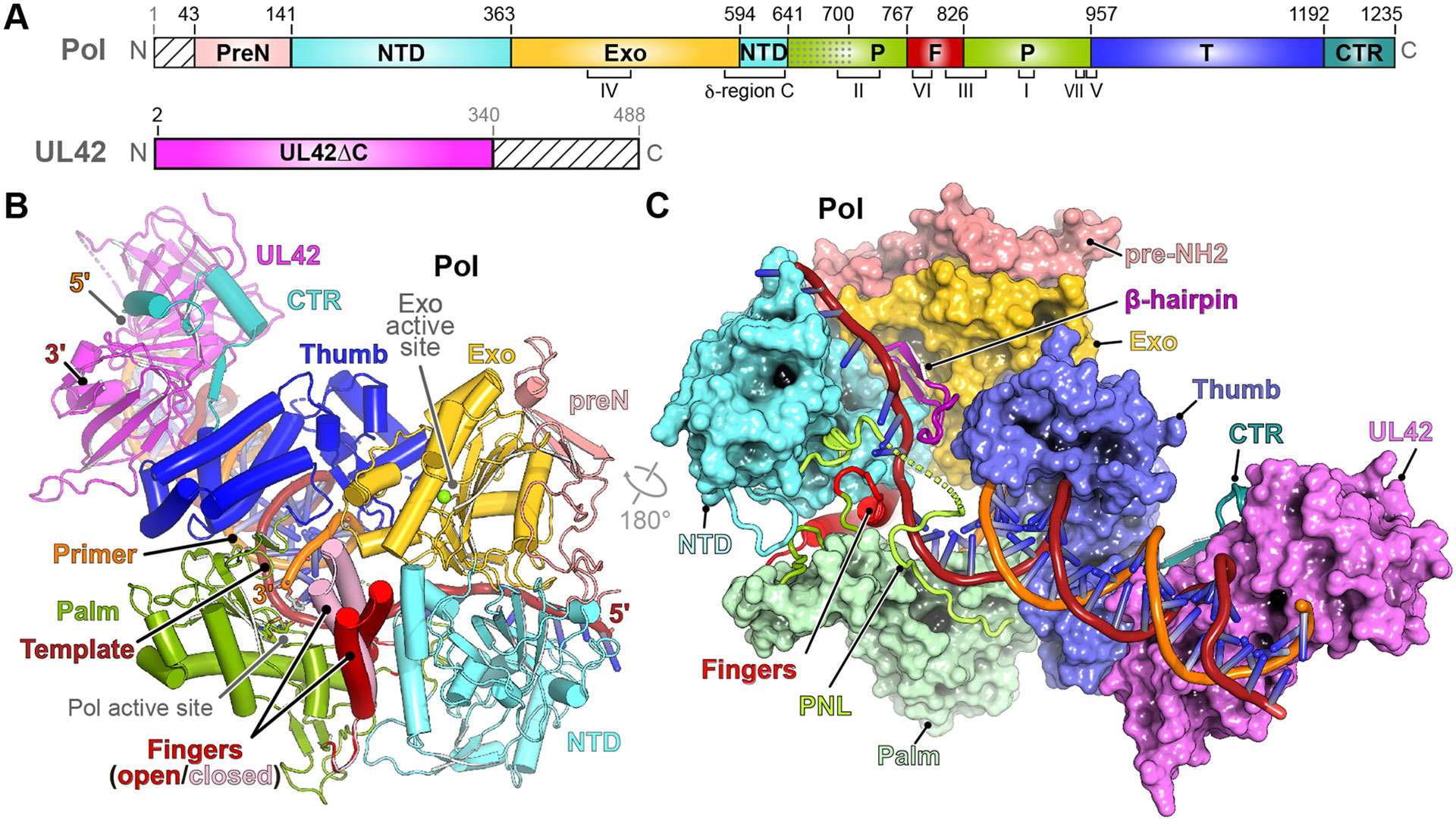
DNA polymerases are important drug targets, and many structural studies have captured them in distinct conformations. However, a detailed understanding of the impact of polymerase conformational dynamics on drug resistance is lacking. We determined cryoelectron microscopy (cryo-EM) structures of DNA-bound herpes simplex virus polymerase holoenzyme in multiple conformations and interacting with antivirals in clinical use. These structures reveal how the catalytic subunit Pol and the processivity factor UL42 bind DNA to promote processive DNA synthesis. Unexpectedly, in the absence of an incoming nucleotide, we observed Pol in multiple conformations with the closed state sampled by the fingers domain. Drug-bound structures reveal how antivirals may selectively bind enzymes that more readily adopt the closed conformation. Molecular dynamics simulations and the cryo-EM structure of a drug-resistant mutant indicate that some resistance mutations modulate conformational dynamics rather than directly impacting drug binding, thus clarifying mechanisms that drive drug selectivity.

Keywords: DNA polymerase; acyclovir; conformational dynamics; cryo-EM; drug resistance; foscarnet; herpesvirus.


Conflict of interest statement

Declaration of interests The authors declare no competing interests.

Figures

Figure 1.
Figure 1.. Structure of the DNA-bound HSV polymerase holoenzyme.
(A) Diagrams of HSV Pol and UL42 constructs used for structural studies. Pol conserved regions (CR) I–VII, are shown in brackets. δ-region C is also indicated. Stripes indicate regions not included in constructs. preN, pre-NH2; NTD, NH2-terminal domain; Exo, exonuclease domain; P, palm; F, fingers; T, thumb; CTR, C-terminal region. The dotted segment indicates the palm-NTD loop. (B) DNA-bound HSV polymerase in the absence of nucleotide. The primer 3' end is in the Pol active site. We observed alternate conformations for the fingers (open and closed) (see Figure S1D). (C) Surface representation of DNA-bound HSV polymerase in a rotated view. The fingers, loops near the base of the fingers, palm-NTD loop, Exo β-hairpin motif, Pol CTR, and DNA are shown as ribbons.
Figure 2.
Figure 2.. HSV Pol palm, palm-NTD loop, and thumb DNA contacts.
(A) Sequence alignment showing the palm-NTD loop (PNL) region (residues 641–700). Red box indicates residues that are disordered in the structure. Within the red box, positively and negatively charged residues are colored in blue and red, respectively; residues with linker-like properties (serine, alanine, glycine, and prolines) are in gray. McHV1: Macacine herpesvirus 1. VZV: Varicella zoster virus. Alignment was generated using ESPript 3.0. (B) PNL DNA contacts for the nucleotide-free polymerase (open conformation). The pseudo base pair-like contacts that the R692Pol side chain makes with the primer −5 base are shown as yellow dashes and other polar contacts with neighboring bases are shown as thin black dashes. Additional side chain and protein backbone contacts involving Pol residues in this region are shown as yellow dashes. (C) Pol palm, thumb, and PNL contacts with DNA for the closed (dTTP-bound) polymerase. CRV and CRVII engage the primer near its 3' end, and the PNL engages the backbone of the DNA template on the opposite side of the duplex. Selected contacts are shown, with polar contacts shown as yellow dashes. See also Fig. S2. In panels B and C, R842Pol, Y941Pol, and N961Pol, indicated by asterisks, are sites of mutations associated with antiviral resistance (see text).
Figure 3.
Figure 3.. DNA-bound HSV polymerase in the closed conformation and active site recognition of nucleotide or antivirals.
(A) HSV polymerase in the closed conformation bound to DNA primer-template with deoxythymidine triphosphate (dTTP) opposite the templating base. dTTP is shown as sticks. (B) HSV Pol active site with dTTP. (C) HSV Pol active site with acyclovir triphosphate (ACV-TP). (D) HSV Pol active site with foscarnet. The primer 3' end is untranslocated. The complexes included a 3'-dideoxy-terminated primer to prevent nucleotide incorporation. Metals are shown as green spheres. In B–D, selected contacts are shown as yellow dashes.
Figure 4.
Figure 4.. Drug-resistance mutations influence polymerase fingers dynamics.
(A) Substitutions that confer resistance to acyclovir and/or a pyrophosphate analog as demonstrated by genetic experiments such as marker transfer are mapped on DNA-bound HSV polymerase in the open conformation. Substituted residues are shown as sticks and colored according to domains. Substitutions and their effects on drug resistance are reviewed in Piret et al., 2021. (B–C) Per-residue root-mean-square-fluctuation (RMSF) from (MD) simulations of the polymerase in the open conformation for wild-type (WT) (blue trace) and the W781V mutant (pink trace) (B), or of the polymerase in the closed conformation for the WT (brown trace) and the W781V mutant (green trace) (C). (D) Mean RMSF values calculated during MD simulations for fingers residues (S772–G822) are plotted for the WT (cryo-EM) and W781V (modeled) structures in the open (top panel) and closed (bottom panel) conformations. (E) View of fingers domain residue W781 and neighboring hydrophobic residues in the open and closed (dTTP-bound) conformations in the cryo-EM structures. (F) View of representative frames from MD simulations to show neighboring hydrophobic residues for the W781V mutant enzyme in the open and closed conformations. (G) Per-residue fluctuation from MD simulations of the holoenzyme in the closed conformation for the WT holoenzyme (top panel) or W781V Pol mutant (bottom panel) in the presence or absence of dTTP. Only the fingers residues are shown. (H) Mean RMSF values calculated during MD simulations for fingers residues (S772–G822) are plotted for the WT (top panel) and W781V mutant (bottom panel) in the closed conformation. For panels B and C, the right panels show plots of the fingers region, indicated by the dashed box in the left panels. Regions denoted “Δ” are absent in models (lacked interpretable density in cryo-EM maps). For panels in G, full tracings are provided in Figure S6. For panels D and H, comparison between two groups was performed using an unpaired, two-tailed Student’s t-test. Error bars represent standard errors. **p <0.01, ***p <0.001.
Figure 5.
Figure 5.. Structure of an editing HSV polymerase.
(A) HSV polymerase bound to DNA primer-template containing a mismatch at the primer 3' end. The fingers are open, and the primer 3' end is translocated into the Exo active site, which is 40 Å away from the Pol active site. (B) Surface representation of the DNA-bound HSV polymerase in the editing conformation in a rotated view. The fingers, loops near the base of the fingers, palm-NTD loop (PNL), Exo β-hairpin motif, and DNA are shown as ribbon diagrams. (C–D) Exo β-hairpin motif and palm-NTD loop interactions in the closed (C) and editing (D) polymerases. For the closed structure (C), the nucleotide residues that are labeled with asterisks were modeled but not deposited in structural coordinates due to weak density. For the editing structure (D), the nucleotide residue at the 3' end of the primer (labeled with an asterisk) and one of the two Exo active site metals (see Figure S7B) were also modeled but not deposited in structural coordinates because of weak density.
Figure 6.
Figure 6.. UL42 DNA binding and comparison to human PCNA.
(A) Electrostatic surface potential of HSV polymerase bound to DNA in the closed conformation (bound to dTTP). DNA primer and template are shown as a ribbon diagram. The Pol thumb and UL42 surfaces are outlined. (B) Closeup view of the UL42-DNA interface highlighting Pol thumb and UL42 residues that contact DNA (<4 Å). (C) Electrostatic surface potential representation of UL42 bound to DNA as part of the HSV polymerase. DNA is shown as a ribbon diagram, and bound nucleotide in the active site is shown as sticks. The catalytic subunit (Pol) is omitted for clarity. Top and bottom panels are different views. (D) Electrostatic surface potential representation of the PCNA trimer surrounding DNA as part of human polymerase δ (PDB ID: 6TNY). The DNA is shown as a ribbon diagram, and bound nucleotide in the active site is shown as sticks. Other subunits (e.g., catalytic and accessory subunits) are omitted for clarity. Top and bottom panels are different views. The positions of two PCNA residues (N84 and R149) that contact the DNA phosphate backbone (<4 Å) are indicated. (E–F) Pol thumb (E) or Pol thumb and CTR (F) contacts with UL42. Contacts shown were not previously visualized in the structure of UL42 bound to Pol CTR residues 1200–1235.

References

    1. Coen DM, Namchuk MN, and Kuritzkes DR (2023). Antiviral agents. In Fields Virology, Howley PM, Knipe DM, Whelan SM, Cohen JI, Damania B, and Freed EO, eds. (Wolters Kluwer; ), pp. 353–397.
    1. Piret J, and Boivin G (2021). DNA polymerases of herpesviruses and their inhibitors. Enzymes 50, 79–132. 10.1016/bs.enz.2021.07.003. - DOI - PubMed
    1. Gottlieb J, Marcy AI, Coen DM, and Challberg MD (1990). The herpes simplex virus type 1 UL42 gene product: a subunit of DNA polymerase that functions to increase processivity. J Virol 64, 5976–5987. - PMC - PubMed
    1. Liu S, Knafels JD, Chang JS, Waszak GA, Baldwin ET, Deibel MR Jr., Thomsen DR, Homa FL, Wells PA, Tory MC, et al. (2006). Crystal structure of the herpes simplex virus 1 DNA polymerase. J Biol Chem 281, 18193–18200. 10.1074/jbc.M602414200. - DOI - PubMed
    1. Zuccola HJ, Filman DJ, Coen DM, and Hogle JM (2000). The crystal structure of an unusual processivity factor, herpes simplex virus UL42, bound to the C terminus of its cognate polymerase. Mol Cell 5, 267–278. 10.1016/s1097-2765(00)80422-0. - DOI - PubMed

MeSH terms