. 2024 Aug 28:15910199241267312.
doi: 10.1177/15910199241267312. Online ahead of print.

Quantifying extent of meningioma preoperative embolization through volumetric analysis: A retrospective case series

Affiliations

Quantifying extent of meningioma preoperative embolization through volumetric analysis: A retrospective case series

Denzel E Faulkner et al. Interv Neuroradiol. .

Abstract

Background: Endovascular embolization is an adjunct to meningioma resection. Isolating the effectiveness of embolization is difficult as MR imaging is typically performed before embolization and after resection, and volumetric assessment of embolization on 2D angiographic imaging is challenging. We investigated the correlation between 2D angiographic and 3D MR measurements of meningioma devascularization following embolization.

Methods: We implemented a protocol for postembolization, preresection MRI. Angiographic devascularization was graded according to reduction of tumor blush from 1 (partial embolization) to 4 (complete embolization with no residual circulation supply). Volumetric extent of embolization was quantified as the percent of tumor contrast enhancement lost following embolization. Tumor embolization was analyzed according to tumor location and vascular supply.

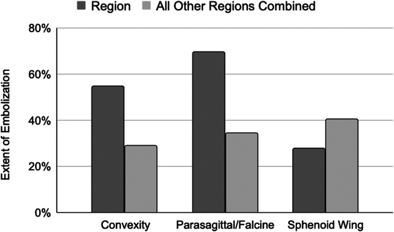
Results: Thirty consecutive patients met inclusionary criteria. Grade 1 devascularization was achieved in 7% of patients, grade 2 in 43%, grade 3 in 20%, and grade 4 in 30%. Average extent of embolization was 37 ± 6%. Extent of tumor embolization was low (<25%) in 40%, moderate (25%-75%) in 40%, and high (>75%) in 20% of patients. Convexity, parasagittal/falcine and sphenoid wing tumors were found to have distinct vascular supply patterns and extent of embolization. Angiographic devascularization grade was significantly correlated with volumetric extent of tumor embolization (p < 0.001, r = 0.758).

Conclusion: This is the first study to implement postembolization, preoperative MRI to assess extent of embolization prior to meningioma resection. The study demonstrates that volumetric assessment of contrast reduction following embolization provides a quantitative and spatially resolved framework for assessing extent of tumor embolization.

Keywords: Tumor embolization; image segmentation; meningioma; volumetric analysis.


Conflict of interest statement

Declaration of conflicting interestsThe author(s) declared no potential conflicts of interest with respect to the research, authorship, and/or publication of this article.

Figures

Figure 1.
Figure 1.
Preparation embolization and resection of a right frontal convexity meningioma. Far Left Column: Contrast-enhanced T1-weighted MR images in the axial (a), sagittal (b), and coronal (c) planes demonstrate an avidly and homogeneously enhancing right frontal convexity meningioma at the time of presentation. Middle-Left Column: A lateral angiogram demonstrated that the tumor was supplied entirely by the right middle meningeal artery (d). The tumor was completely embolized with n-BCA glue via the right middle meningeal artery, and the postembolization angiogram demonstrated no residual tumor blush (e). Middle-Right Column: Postembolization contrast-enhanced T1-weighted MR images in the axial (g), sagittal (h), and coronal (i) planes demonstrate near-complete loss of contrast enhancement relative to the corresponding preembolization images. At the time of resection, one day after embolization, the tumor was found to be completely devascularized, necrotic, and soft (f). The tumor was removed with minimal blood loss. Far Right Column: The immediate postoperative images corresponding to those in the other columns are shown in the axial (g), sagittal (h), and coronal (i) planes. These contrast-enhanced T1-weighted MR images demonstrate gross total resection.
Figure 2.
Figure 2.
Tumor segmentation for volumetric analysis before and after embolization. Magnetic resonance imaging (MRI) scans of the brain in a patient with a left lateral sphenoid wing meningioma before and after embolization (a–f), and with color-coding of the tumor segmentation used for volumetric analysis (g–l). All images are derived from contrast-enhanced T1 sequences. Far Left Column: Preembolization (a) axial, (b) sagittal, and (c) coronal images. Middle-Left Column: Postembolization, preresection (d) axial, (e) sagittal, and (f) coronal images. Middle-Right Column: Color-coded preembolization (g) axial, (h) sagittal, and (i) coronal images. Far Right Column: Color-coded postembolization (j) axial, (k) sagittal, and (l) coronal images. The magenta volumes reflect regions of contrast enhancement within the tumor at each time point. The green volumes represent regions of nonenhancing tumor. Extent of tumor embolization in this case was ε = 82%.
Figure 3.
Figure 3.
Extent of tumor embolization in key locations in contrast to all other tumor locations. Extent of tumor embolization is compared for the key tumor locations discussed in the text (parasagittal, convexity, and sphenoid wing). These are contrasted with the average extent of tumor embolization of all other groups and a two sample t-test was conducted for all pairs. Tumors in the parasagittal/falcine and convexity location had higher likelihood of having a high and moderate extent of embolization, respectively, whereas sphenoid wing tumors were likely to have lower extent of embolization as compared to other regions. ** The p value for these trends was greater than 0.05.
Figure 4.
Figure 4.
Relationship between extent of embolization and devascularization grading. Extent of tumor embolization as assessed by MRI using the methods described here corresponds closely to the devascularization grade obtained through angiographic assessment.

Similar articles

Cited by

References

    1. Wei Z, Mallela AN, Faramand A, et al. Long-term survival in patients with long-segment complex meningiomas occluding the dural venous sinuses: illustrative cases. J Neurosurg Case Lessons Epub ahead of print 17 May 2021; 1. - PMC - PubMed
    1. Huntoon K, Toland AMS, Dahiya S. Meningioma: a review of clinicopathological and molecular aspects. Front Oncol 2020; 10: 579599. - PMC - PubMed
    1. Gruber P, Schwyzer L, Klinger E, et al. Longitudinal imaging of tumor volume, diffusivity, and perfusion after preoperative endovascular embolization in supratentorial hemispheric meningiomas. World Neurosurg 2018; 120: e357–e364. - PubMed
    1. Matsoukas S, Feng R, Gilligan J, et al. Combined transarterial and percutaneous preoperative embolization of transosseous meningioma. Interv Neuroradiol 2023; 29: 618. - PMC - PubMed
    1. Schartz D, Manganaro M, Szekeres D, et al. Direct percutaneous puncture versus transarterial embolization for head and neck paragangliomas: a systematic review and meta-analysis. Interv Neuroradiol 2023; 29: 15910199231188859. - PubMed

LinkOut - more resources