. 2024 Aug 28;11(1):940.
doi: 10.1038/s41597-024-03775-2.

High-resolution segmentations of the hypothalamus and its subregions for training of segmentation models

Affiliations

High-resolution segmentations of the hypothalamus and its subregions for training of segmentation models

Livia Rodrigues et al. Sci Data. .

Abstract

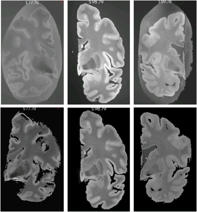
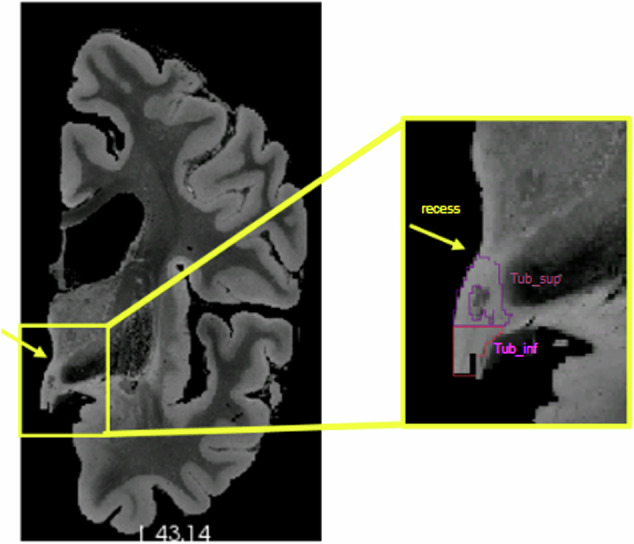
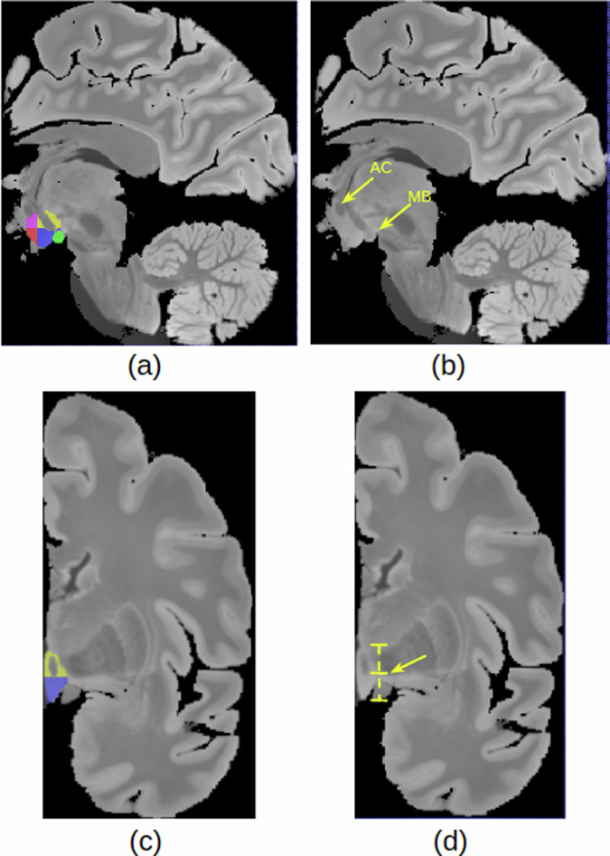
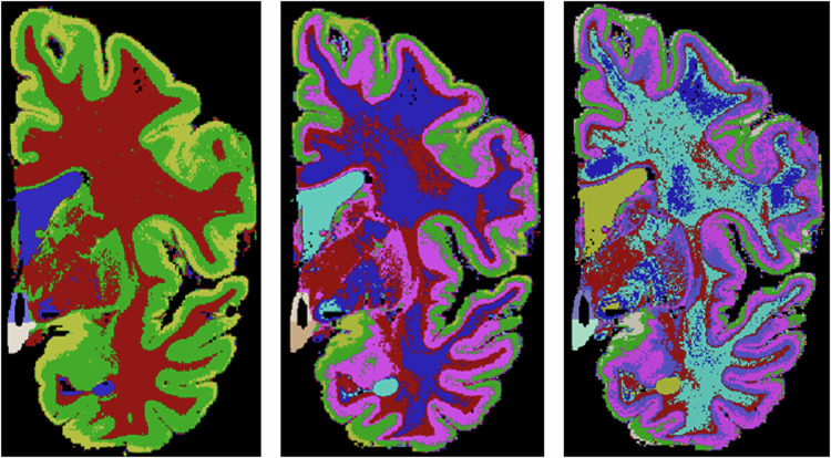
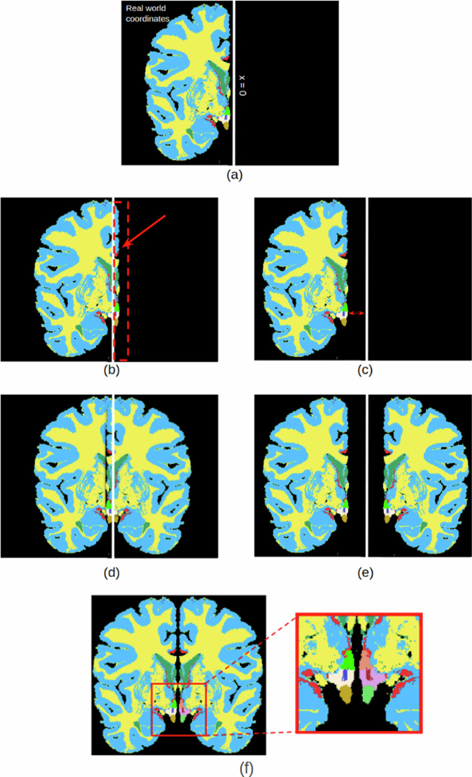
Segmentation of brain structures on magnetic resonance imaging (MRI) is a highly relevant neuroimaging topic, as it is a prerequisite for different analyses such as volumetry or shape analysis. Automated segmentation facilitates the study of brain structures in larger cohorts when compared with manual segmentation, which is time-consuming. However, the development of most automated methods relies on large and manually annotated datasets, which limits the generalizability of these methods. Recently, new techniques using synthetic images have emerged, reducing the need for manual annotation. Here we provide a dataset composed of label maps built from publicly available ultra-high resolution ex vivo MRI from 10 whole hemispheres, which can be used to develop segmentation methods using synthetic data. The label maps are obtained with a combination of manual labels for the hypothalamic regions and automated segmentations for the rest of the brain, and mirrored to simulate entire brains. We also provide the pre-processed ex vivo scans, as this dataset can support future projects to include other structures after these are manually segmented.


Conflict of interest statement

The authors declare no competing interests.

Figures

Fig. 1
Fig. 1
Examples of original ex vivo images (up) and images after the pre-processing steps (down). First, we re-orient the images to positive RAS standard and remove non-cerebral elements from the background. Then, we resample the images voxels to 0.3 × 0.3 × 0.3 mm isotropic and perform bias field correction.
Fig. 2
Fig. 2
Recess of the hypothalamus used for the delineation of superior boundary. Tub_sup = Tuberal Superior subregion;Tub_inf = Tuberal inferior subregion.
Fig. 3
Fig. 3
Segmentation protocol: (a) Subregions delineation: Anterior superior (pink), anterior inferior (red), tuberal superior (yellow), tuberal inferior (blue) and posterior (green) (b)Sagittal landmarks for subregion delineation (AC = Anterior Comissure and MB=mamillary body) (c) Coronal View of superior and inferior tuberal subregions (d) Coronal landmark for superior/inferior division.
Fig. 4
Fig. 4
Examples of three different label maps derived from the same image. From left to right: k = 4, k = 6, k = 8.
Fig. 5
Fig. 5
(a) Whole brain segmentation: example with k = 4 (b) Manual segmentation simply overlapping the whole brain segmentation. We can see that there are a few inconsistent voxels, that should be labeled either as hypothalamus or background that have different labels. (c) To fix these inconsistencies, a mathematical morphology-based algorithm is applied.
Fig. 6
Fig. 6
Label map creation: Following the segmentation step, a half-brain label map is generated (a). However, given the hypothalamus’s central location within the brain, mirroring is essential to provide contextual information. For the mirroring process, translation and rotation are applied to the RAS coordinates. This involves moving the brain close to the x = 0 axis from the negative side, without surpassing into the positive side. Essentially, a final cost function is computed, penalizing positive values of x (b) and high negative values. Finally, we prevent the overlap between brain hemispheres (d) and also prevent them from ending up at unnaturally distant positions (e). After the optimization and mirroring, we end up with the final label map (f).
Fig. 7
Fig. 7
Hypothalamus and subregions segmentation on in vivo images using the method trained on the synthetic dataset. The method was capable of segmenting the hypothalamus in T1w, T2w, PD, and FLAIR sequences, the last one presenting a spacing of 5 mm.

References

    1. Saper, C. B. & Lowell, B. B. The hypothalamus. Current Biology24, R1111–R1116 (2014). 10.1016/j.cub.2014.10.023 - DOI - PubMed
    1. Neudorfer, C. et al. A high-resolution in vivo magnetic resonance imaging atlas of the human hypothalamic region. Scientific Data7, 305 (2020). 10.1038/s41597-020-00644-6 - DOI - PMC - PubMed
    1. Vercruysse, P. & Vieau, D. et al. Hypothalamic alterations in neurodegenerative diseases and their relation to abnormal energy metabolism. Front.Mol. Neurosci.11, 2 (2018). 10.3389/fnmol.2018.00002 - DOI - PMC - PubMed
    1. Piyush, R. & Ramakrishnan, S. Analysis of sub-anatomic volume changes in Alzheimer brain using diffusion tensor imaging. In 2014 40th Annual Northeast Bioengineering Conference (NEBEC), 1–2 (IEEE, 2014).
    1. Gabery, S. et al. Volumetric analysis of the hypothalamus in huntington disease using 3T MRI: The image-hd study. PloS one10, e0117593 (2015). 10.1371/journal.pone.0117593 - DOI - PMC - PubMed

LinkOut - more resources