Review
. 2025 Feb;22(2):90-104.
doi: 10.1038/s41569-024-01067-1. Epub 2024 Aug 28.

Pathophysiological insights into HFpEF from studies of human cardiac tissue

Affiliations
Review

Pathophysiological insights into HFpEF from studies of human cardiac tissue

Ahmed U Fayyaz et al. Nat Rev Cardiol. 2025 Feb.

Abstract

Heart failure with preserved ejection fraction (HFpEF) is a major, worldwide health-care problem. Few therapies for HFpEF exist because the pathophysiology of this condition is poorly defined and, increasingly, postulated to be diverse. Although perturbations in other organs contribute to the clinical profile in HFpEF, altered cardiac structure, function or both are the primary causes of this heart failure syndrome. Therefore, studying myocardial tissue is fundamental to improve pathophysiological insights and therapeutic discovery in HFpEF. Most studies of myocardial changes in HFpEF have relied on cardiac tissue from animal models without (or with limited) confirmatory studies in human cardiac tissue. Animal models of HFpEF have evolved based on theoretical HFpEF aetiologies, but these models might not reflect the complex pathophysiology of human HFpEF. The focus of this Review is the pathophysiological insights gained from studies of human HFpEF myocardium. We outline the rationale for these studies, the challenges and opportunities in obtaining myocardial tissue from patients with HFpEF and relevant comparator groups, the analytical approaches, the pathophysiological insights gained to date and the remaining knowledge gaps. Our objective is to provide a roadmap for future studies of cardiac tissue from diverse cohorts of patients with HFpEF, coupling discovery biology with measures to account for pathophysiological diversity.


Conflict of interest statement

Competing interests: The authors declare no competing interests.

Figures

Fig. 1 |
Fig. 1 |. Structural, functional and metabolic alterations in the myocardium from patients with HFpEF.
Myocardial fibrosis, cardiomyocyte hypertrophy, microvascular rarefaction, transverse tubule (T-tubule) dilatation and proliferation, impaired calcium homeostasis, titin hypophosphorylation, prolonged cardiomyocyte relaxation, increased cardiomyocyte stiffness (data from ref. 122), increased passive force (data from ref. 60), and metabolic impairment have been demonstrated in the myocardium from patients with heart failure with preserved ejection fraction (HFpEF). RYR2, ryanodine receptor 2; SERCA2, sarcoplasmic–endoplasmic reticulum calcium ATPase 2; TCA, tricarboxylic acid.
Fig. 2 |
Fig. 2 |. Pathophysiological mechanisms in human HFpEF.
Pathophysiological mechanisms involved in heart failure with preserved ejection fraction (HFpEF) based on findings reported in studies using human myocardial samples. a, Natriuretic peptides (NPs), via particulate guanylyl cyclase (pGC), and nitric oxide (NO), via soluble guanylyl cyclase (sGC), stimulate the production of cGMP from GTP, which in turn stimulates the activation of cGMP-dependent protein kinase (also known as PKG). High-affinity cGMP-specific 3′,5′-cyclic phosphodiesterase type 9A (PDE9A) and phosphodiesterase 5 (PDE5) degrade NP-derived and NO-derived cGMP, respectively. Activation of the cGMP–PKG pathway is believed to prevent or reduce myocardial hypertrophy and fibrosis, enhance myocardial relaxation (lusitropy), reduce myocardial stiffness (via phosphorylation of titin) and promote clearance of dysfunctional proteins. In human HFpEF myocardium, evidence of downregulation of this pathway includes reductions in sGC activity, NO concentration and PKG activity, and elevated PDE9 levels. b, Cardiac inflammation is believed to reduce endothelial NO synthase (eNOS) but increase inducible NO synthase (iNOS), and increase oxidative stress, fibrosis, hypertrophy and mitochondrial dysfunction. In human HFpEF, evidence of myocardial inflammation includes increased levels of viral genetic material (indicative of past viral myocarditis) in some patients and increased presence of inflammatory cells, cytokines (tumour necrosis factor (TNF), IL-1 and IL-6), intercellular adhesion molecules (ICAM) and vascular cell adhesion protein 1 (VCAM1). c, Risk factors for HFpEF might be associated with impaired protein synthesis, folding and transport in the endoplasmic reticulum (ER), with accumulation of misfolded proteins (ER stress). ER stress ultimately results in impaired protein quality and cell death. ER stress triggers the unfolded protein response (UPR), which includes three pathways to mitigate ER stress, maintain protein quality control and restore cellular homeostasis and cardiomyocyte function. In human HFpEF, evidence of impaired UPR includes increased levels of iNOS, decreased phosphorylation of serine/threonine protein kinase–endoribonuclease IRE1 and decreased levels of properly spliced XBP1 mRNA (XBP1s), which encodes X-box-binding protein 1 (XBP1). These findings are attributed to inflammation-induced iNOS activation with S-nitrosylation of IRE1, which prevents IRE1 phosphorylation. The reduction in XBP1s mRNA is predicted to limit XBP1 protein production and downstream UPR signalling. d, Environmental agents, reactive oxygen species generated by inflammation and other mechanisms can damage DNA, impair transcription and replication, and lead to impaired cell function. DNA damage activates serine protein kinase ATM (also known as ataxia–telangiectasia mutated kinase), serine protein kinase ATR (also known as ataxia telangiectasia and Rad3-related kinase) and other DNA damage mediators as well as the downstream serine/threonine protein kinases CHK1 and CHK2, which activate DNA damage response effectors, such as cellular tumour antigen p53 and dual specificity phosphatase CDC25. This response can either repair the DNA damage or not, with unrepaired DNA damage leading to cellular senescence and dysfunction. In human HFpEF, evidence of increased DNA damage response as a pathological mechanism includes increases in DNA damage response markers (phosphorylated histone variant H2AX (γH2AX) and the phosphorylated forms of CHK1 and CHK2). TRAF2, TNF receptor-associated factor 2.
Fig. 3 |
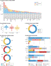
Fig. 3 |. Studies of tissue samples from patients with HFpEF and comparators.
a, Number of patients per group in each study,–,,,,,–. b, Mean or median age of patients by group. c, Ejection fraction criteria for heart failure with preserved ejection fraction (HFpEF) designation. d, Frequency of tissue acquisition methods. e, Sites from which cardiac tissue was harvested and number of studies for each site. The ‘other’ sites include mixtures of samples from the right and left sides of the heart, right atrial (RA) tissue, epicardial adipose tissue or a mix of RA and left ventricular (LV) tissues or unspecified LV site samples. EMB, endomyocardial biopsy; HFrEF, heart failure with reduced ejection fraction; NF, non-failing (without heart failure); RV, right ventricular.

Similar articles

Cited by

References

    1. Savarese G et al. Global burden of heart failure: a comprehensive and updated review of epidemiology. Cardiovasc Res 118, 3272–3287 (2022). - PubMed
    1. Borlaug BA, Sharma K, Shah SJ & Ho JE Heart failure with preserved ejection fraction: JACC scientific statement. J. Am. Coll. Cardiol 81, 1810–1834 (2023). - PubMed
    1. Dunlay SM, Roger VL & Redfield MM Epidemiology of heart failure with preserved ejection fraction. Nat. Rev. Cardiol 14, 591–602 (2017). - PubMed
    1. Redfield MM & Borlaug BA Heart failure with preserved ejection fraction: a review. JAMA 329, 827–838 (2023). - PubMed
    1. Desai AS, Lam CSP, McMurray JJV & Redfield MM How to manage heart failure with preserved ejection fraction: practical guidance for clinicians. JACC Heart Fail 11, 619–636 (2023). - PubMed

LinkOut - more resources