. 2024 Jul 29;13(8):914.
doi: 10.3390/antiox13080914.

Evidence for TGF-β1/Nrf2 Signaling Crosstalk in a Cuprizone Model of Multiple Sclerosis

Affiliations

Evidence for TGF-β1/Nrf2 Signaling Crosstalk in a Cuprizone Model of Multiple Sclerosis

Coram Guevara et al. Antioxidants (Basel). .

Abstract

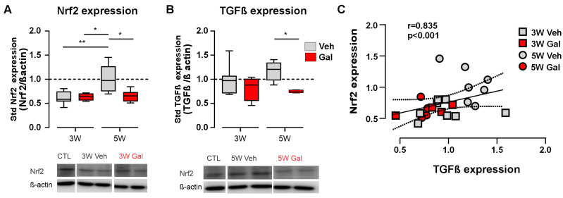
Multiple sclerosis (MS) is a chronic and degenerative disease that impacts central nervous system (CNS) function. One of the major characteristics of the disease is the presence of regions lacking myelin and an oxidative and inflammatory environment. TGF-β1 and Nrf2 proteins play a fundamental role in different oxidative/inflammatory processes linked to neurodegenerative diseases such as MS. The evidence from different experimental settings has demonstrated a TGF-β1-Nrf2 signaling crosstalk under pathological conditions. However, this possibility has not been explored in experimental models of MS. Here, by using the cuprizone-induced demyelination model of MS, we report that the in vivo pharmacological blockage of the TGF-β1 receptor reduced Nrf2, catalase, and TGFβ-1 protein levels in the demyelination phase of cuprizone administration. In addition, ATP production, locomotor function and cognitive performance were diminished by the treatment. Altogether, our results provide evidence for a crosstalk between TGF-β1 and Nrf2 signaling pathways under CNS demyelination, highlighting the importance of the antioxidant cellular response of neurodegenerative diseases such as MS.

Keywords: Nrf2; demyelination; multiple sclerosis; neuroinflammation; oxidative stress; transforming growth factor β1 TGF-β1.


Conflict of interest statement

The authors declare no conflicts of interest.

Figures

Figure 1
Figure 1
Experimental model of MS with cuprizone-induced demyelinated lesions. (A) Wild-type mice were fed cuprizone (CPZ, 0.25%) for 5 weeks. The treated group received the TGF-β1 receptor blocker galunisertib (GAL, 10 mg/kg) daily during the third week of CPZ feeding. Note the expected reduction in pSMAD in the mice treated with the TGF-β1 receptor blocker GAL compared to the control animals (B) (*) p < 0.05. (C) Immunofluorescence of 75-µm-thick callosal coronal sections from the control mice or mice fed with 0.25% cuprizone over 3 and 5 weeks (CPZ 3W and 5W, respectively). Dotted lines indicate the corpus callosum (CC). Demyelinated areas, recognized by the lack of myelin basic protein (MBP) expression (blue), are indicated by red asterisks. The summary of the normalized lesion size is also shown (plot, Mann–Whitney test, (**) p < 0.01, n = 3–4 animals). (D) Protein levels for the oligodendroglial marker OLG2 at basal line (Ctl) and weeks 5 and 13 (CPZ 5W and 13W, respectively) of the cuprizone model (Kruskal–Wallis test, (**) p < 0.01, n = 3–4; representative images of western blot membranes from two mice are shown).
Figure 2
Figure 2
TGF-β1 receptor 1 blocking reduces both Nrf2 and TGF-β1 protein levels in the CPZ model of MS. (A,B) Protein levels in respect to the control and normalized by β-actin for Nrf2 (A) and TGF-β1 (B) in the corpus callosum of the mice fed with CPZ over 3 or 5 weeks (3W and 5W) treated with galunisertib (GAL, 10 mg/kg, red blocks) or vehicle (Veh, gray blocks). Representative images of western blot membranes from two mice are shown (bottom). Kruskal–Wallis test, Dunn’s multiple comparison post hoc tests (*) p > 0.05, (**), p < 0.01. (n = 4–8 mice). (C) Correlation between protein levels for TGF-β1 and Nrf2 at the different time points and treatments (r = 0.835, p < 0.001, Spearman test).
Figure 3
Figure 3
TGF-β1 receptor 1 blocking reduces both ATP production and catalase protein levels in the CPZ model of MS. (A) ATP bioluminescence quantification in the corpus callosum of the mice fed with CPZ over 3 or 5 weeks (3W and 5W) treated with galunisertib (GAL, 10 mg/kg, red blocks) or vehicle (Veh, gray blocks). (B) Protein levels in respect to the control and normalized by β-actin for catalase (CAT) in the corpus callosum of the mice fed with CPZ over 3 or 5 weeks (3W and 5W) treated with galunisertib (GAL, 10 mg/kg, red blocks) or vehicle (Veh, gray blocks). (C) Representative images of western blot membranes from two mice are shown. Data in A are expressed as the mean ± standard error. Kruskal–Wallis test, Dunn’s multiple comparison post hoc tests (*) p < 0.05; (**), p < 0.01: (***), p < 0001. (n = 3–6 mice for ATP measurements, n = 4–6 mice for CAT levels).
Figure 4
Figure 4
TGF-β1 receptor 1 blocking reduces the locomotor performance and the cognitive function in the CPZ animal model of MS. (A) Summary of the standardized time-to-fall in the rotarod apparatus for mice before (basal) and after being fed with CPZ for 5 weeks with no treatment (control, Ctl, dark-gray blocks) or treated with galunisertib (GAL, 10 mg/Kg, red blocks) or vehicle (Veh, light-gray blocks). Note that all data are normalized in respect to the basal value with no treatment. (B) Summary of the interaction (D2) index with the novel object for mice before (basal) and after being fed with CPZ for 5 weeks with no treatment (control, Ctl, dark-gray blocks) or treated with galunisertib (GAL, 10 mg/Kg, red blocks) or vehicle (Veh, light-gray blocks). Kruskal–Wallis test, Dunn’s multiple comparison post hoc tests, (*) p < 0.05 (n = 3–8 mice for locomotor test, n = 3–7 mice for NOR test).
Figure 5
Figure 5
Putative mechanisms of the TGF-β1/Nrf2 signaling crosstalk in demyelinated lesions. Under demyelination by cuprizone, TGF-β1 expression increases, most likely promoting astrocyte and microglia TGF-β1 release. The activation of the TGF-β1 receptor will, in turn, activate an Nrf2-dependent antioxidant response, such as enhanced catalase (CAT) expression and ATP synthesis. Thus, blocking the TGF-β1 receptor reduces this response by mechanisms to be determined (?), triggering a failure in antioxidant-related recovery (for instance, locomotor and cognitive function). This figure was created in Biorender.

References

    1. Dobson R., Giovannoni G. Multiple Sclerosis—A Review. Eur. J. Neurol. 2019;26:27–40. doi: 10.1111/ene.13819. - DOI - PubMed
    1. Kobelt G., Thompson A., Berg J., Gannedahl M., Eriksson J. New Insights into the Burden and Costs of Multiple Sclerosis in Europe. Mult. Scler. 2017;23:1123–1136. doi: 10.1177/1352458517694432. - DOI - PMC - PubMed
    1. Coyle P.K. Multiple Sclerosis in Pregnancy. Continuum. 2014;20:42–59. doi: 10.1212/01.CON.0000443836.18131.c9. - DOI - PMC - PubMed
    1. Cuevas C., Velázquez M., Núñez L., Skromne E., Árcega R., Barroso N., Alberto C., Felipe G., Victor G., Rafael J., et al. Consenso Mexicano Para La Esclerosis Múltiple. Guía Diagnóstica y Terapéutica. Rev. Mex. Neuroci. 2007;8:155–162.
    1. Popescu B.F.G., Lucchinetti C.F. Pathology of Demyelinating Diseases. Annu. Rev. Pathol. Mech. Dis. 2012;7:185–217. doi: 10.1146/annurev-pathol-011811-132443. - DOI - PubMed

Grants and funding

LinkOut - more resources