. 2024 Aug 17;14(8):1023.
doi: 10.3390/biom14081023.

RNA Surveillance Factor SMG5 Is Essential for Mouse Embryonic Stem Cell Differentiation

Affiliations

RNA Surveillance Factor SMG5 Is Essential for Mouse Embryonic Stem Cell Differentiation

Chengyan Chen et al. Biomolecules. .

Abstract

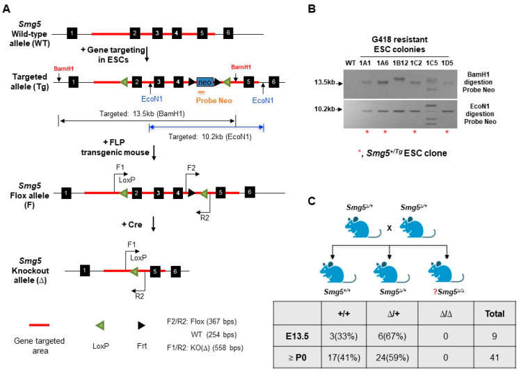
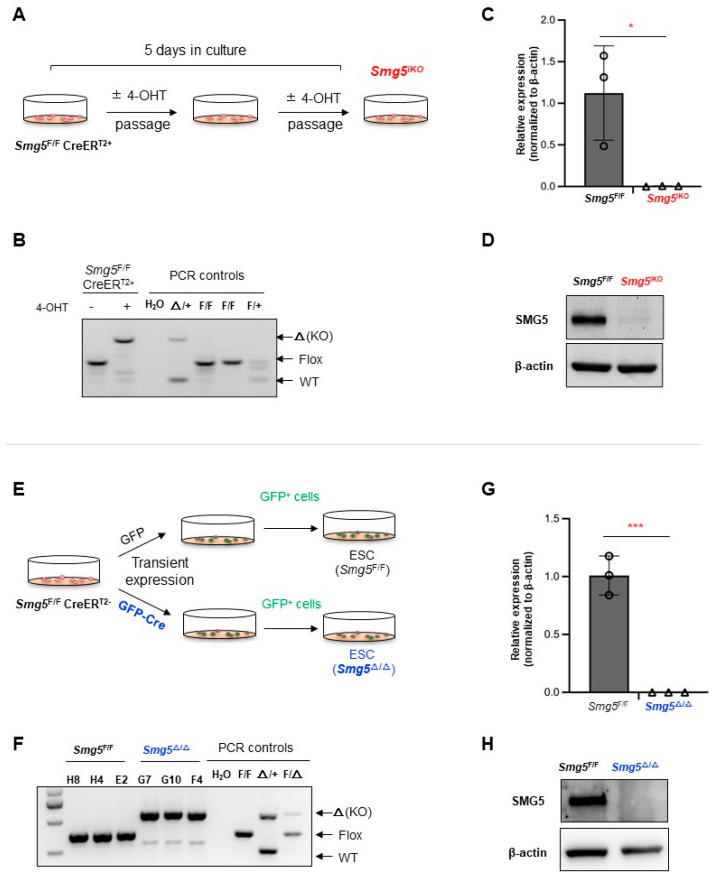
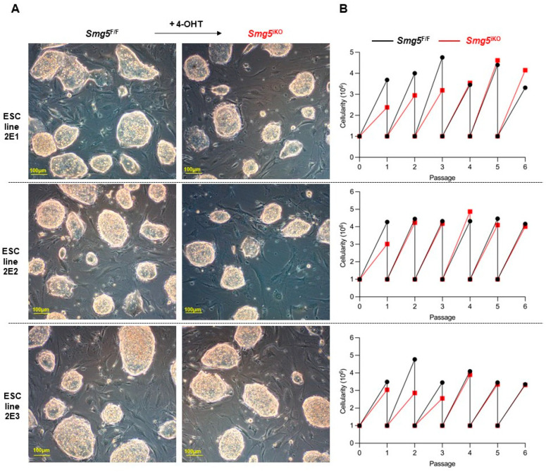
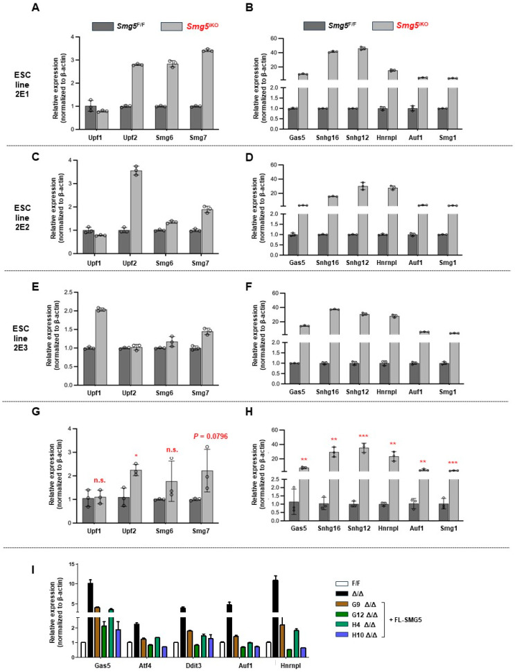
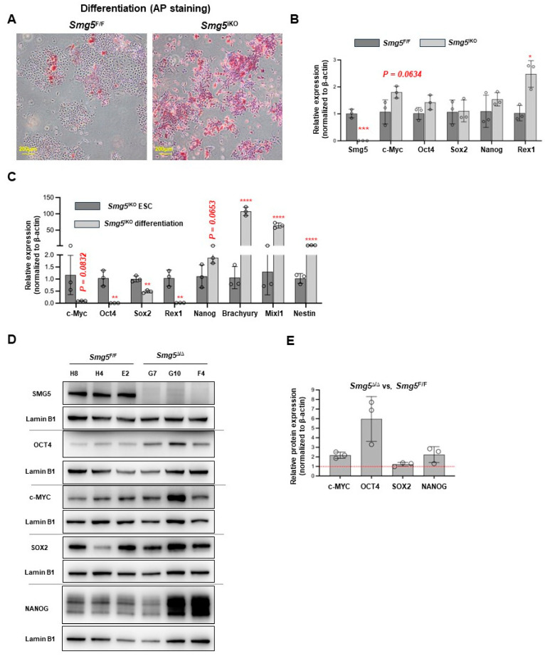
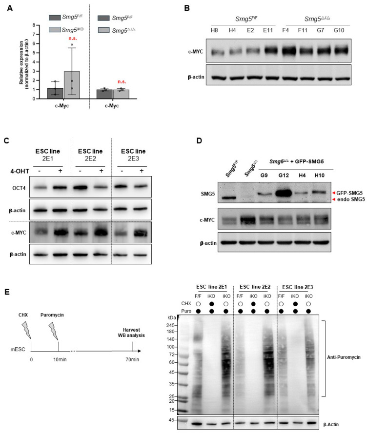
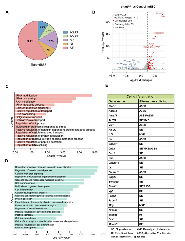
Nonsense-mediated mRNA decay (NMD) is a highly conserved post-transcriptional gene expression regulatory mechanism in eukaryotic cells. NMD eliminates aberrant mRNAs with premature termination codons to surveil transcriptome integrity. Furthermore, NMD fine-tunes gene expression by destabilizing RNAs with specific NMD features. Thus, by controlling the quality and quantity of the transcriptome, NMD plays a vital role in mammalian development, stress response, and tumorigenesis. Deficiencies of NMD factors result in early embryonic lethality, while the underlying mechanisms are poorly understood. SMG5 is a key NMD factor. In this study, we generated an Smg5 conditional knockout mouse model and found that Smg5-null results in early embryonic lethality before E13.5. Furthermore, we produced multiple lines of Smg5 knockout mouse embryonic stem cells (mESCs) and found that the deletion of Smg5 in mESCs does not compromise cell viability. Smg5-null delays differentiation of mESCs. Mechanistically, our study reveals that the c-MYC protein, but not c-Myc mRNA, is upregulated in SMG5-deficient mESCs. The overproduction of c-MYC protein could be caused by enhanced protein synthesis upon SMG5 loss. Furthermore, SMG5-null results in dysregulation of alternative splicing on multiple stem cell differentiation regulators. Overall, our findings underscore the importance of SMG5-NMD in regulating mESC cell-state transition.

Keywords: SMG5; c-MYC; differentiation; mouse embryonic stem cells; nonsense-mediated mRNA decay.


Conflict of interest statement

The authors declare no competing interests.

Figures

Figure 1
Figure 1
Generation of SMG5 inducible knockout mice. (A) Gene targeting strategy to generate Smg5 conditional knockout mice (Smg5F/F). Wild-type allele (WT), gene-targeted allele (Tg), Flox allele (F), and knockout allele (Δ) are shown. Genotyping primers (F1, F2, and R2) are marked on the Smg5 allele. (B) Southern blotting screening of mESC clone selected with G418 (Red asterisks indicate the correct gene-targeted ESC colonies). (C) Mating strategy to generate Smg5Δ/Δ mice and genetic analysis showed that Smg5Δ/Δ mice died before E13.5 (Note: P0, post-natal day 0). Original images can be found in Supplementary Materials.
Figure 2
Figure 2
Establishment of Smg5 deficient mESCs. (A) Strategy for establishing Smg5iKO mESC lines by 4-OHT induction in Smg5F/FCreERT2+ mESCs. (B) PCR analysis on Smg5 locus (exons 2–4) deletion in Smg5F/FCreERT2+ mESCs after 4-OHT treatment for 5 days. (C) qPCR analysis on Smg5 mRNA expression in Smg5F/FCreERT2+ mESCs after 4-OHT treatment for 5 days. (D) Western blotting analysis on SMG5 protein expression in Smg5F/FCreERT2+ mESCs after 4-OHT treatment for 5 days. β-actin is used as a loading control. (E) Strategy to establish Smg5Δ/Δ mESC lines by transfecting pCAG-GFP-Cre into Smg5F/FCreERT2- mESCs. (F) PCR analysis on Smg5 locus (exons 2-4) deletion in Smg5Δ/Δ mESCs. (G) qPCR analysis on Smg5 mRNA expression in Smg5Δ/Δ mESCs. (H) Western blotting analysis on SMG5 protein expression in Smg5Δ/Δ mESCs. β-actin is used as a loading control. Note: unpaired Student’s t-test was carried out for statistical analysis. *, p < 0.05; ***, p < 0.001. Original images can be found in Supplementary Materials.
Figure 3
Figure 3
SMG5 is dispensable for mESC proliferation. (A) Morphology of control and Smg5iKO mESCs on feeders. (B) Proliferation curve of control and Smg5iKO mESCs (6 passages of these mESCs are investigated). Note: 2E1, 2E2, and 2E3 are the parental Smg5 inducible knockout mESC lines.
Figure 4
Figure 4
SMG5 is required for NMD activity in mESCs. (A) qPCR analysis on expressions of NMD factors (Upf1, Upf2, Smg6, and Smg7) in both control and Smg5iKO mESCs (2E1 is the parental Smg5 inducible knockout mESC line). Error bars represent the variance in technical replicates in qPCR assay. (B) qPCR analysis on expressions of NMD canonical targets (Gas5, Snhg16, Snhg12, Hnrnpl, Auf1, and Smg1) in control and Smg5iKO mESCs (2E1 is the parental Smg5 inducible knockout mESC line). Error bars represent the variance of technical replicates in qPCR assay. (C) qPCR analysis on expressions of NMD factors (Upf1, Upf2, Smg6, and Smg7) in both control and Smg5iKO mESCs (2E2 is the parental Smg5 inducible knockout mESC line). Error bars represent the variance in technical replicates in qPCR assay. (D) qPCR analysis on expressions of NMD canonical targets (Gas5, Snhg16, Snhg12, Hnrnpl, Auf1, and Smg1) in control and Smg5iKO mESCs (2E2 is the parental Smg5 inducible knockout mESC line). Error bars represent the variance in technical replicates in qPCR assay. (E) qPCR analysis on expressions of NMD factors (Upf1, Upf2, Smg6, and Smg7) in both control and Smg5iKO mESCs (2E3 is the parental Smg5 inducible knockout mESC line). Error bars represent the variance in technical replicates in qPCR assay. (F) qPCR analysis on expressions of NMD canonical targets (Gas5, Snhg16, Snhg12, Hnrnpl, Auf1, and Smg1) in control and Smg5iKO mESCs (2E3 is the parental Smg5 inducible knockout mESC line). Error bars represent the variance in technical replicates in qPCR assay. (G) qPCR analysis on expressions of NMD factors (Upf1, Upf2, Smg6, and Smg7) in both control and Smg5iKO ESCs (data are summarized from 2E1, 2E2, and 2E3 mESCs). (H) qPCR analysis on expressions of NMD canonical targets (Gas5, Snhg16, Snhg12, Hnrnpl, Auf1, and Smg1) in control and Smg5iKO mESCs (data are summarized from 2E1, 2E2, and 2E3 mESCs). (I) qPCR analysis on expressions of NMD targets in Smg5Δ/Δ mESCs stably expressing GFP-SMG5 fusion protein. Note: unpaired Student’s t-test was carried out for statistical analysis. n.s., not significant; *, p < 0.05; **, p < 0.01; ***, p < 0.001.
Figure 5
Figure 5
Deletion of SMG5 delays mESC differentiation. (A) Alkaline phosphatase (AP) staining on control and Smg5iKO mESCs at differentiation day 4. (B) qPCR analysis on expressions of stemness markers in control and Smg5iKO mESCs (day 4 under spontaneous differentiation condition); (C) qPCR analysis on expressions of stemness markers and lineage makers in Smg5iKO mESCs and Smg5iKO mESCs under spontaneous differentiation condition (day 4). (D) Western blotting analysis on expressions of stemness markers in control and Smg5Δ/Δ mESCs under spontaneous differentiation condition (day 4). Lamin B1 was used as a loading control. (E) Quantification of the protein levels in control and Smg5Δ/Δ mESCs under spontaneous differentiation condition (day 4). Unpaired Student’s t-test was carried out for statistical analysis. *, p < 0.05; **, p < 0.01; ***, p < 0.001; ****, p < 0.0001. Original images can be found in Supplementary Materials.
Figure 6
Figure 6
SMG5 regulates c-MYC expression. (A) qPCR analysis on the expression of c-Myc mRNAs in control, Smg5iKO, and Smg5Δ/Δ mESCs. (B) Western blotting analysis on c-MYC protein expression in control and Smg5Δ/Δ mESCs. (C) Western blotting analysis on expression of c-MYC proteins in control and Smg5iKO ESCs. Note: 2E1, 2E2, and 2E3 are the parental Smg5 inducible knockout mESC lines. (D) Western blotting analysis on c-MYC protein expression in distinct Smg5-deficient mESC clones expressing GFP-SMG5. An SMG5 antibody is used to detect the fusion proteins. (E) SUnSET assay in control and Smg5iKO ESCs. Global protein synthesis (indicated by puromycin incorporation) was revealed by the anti-puromycin antibody staining on control and Smg5iKO mESC protein samples. β-actin is used as a loading control for Western blotting. Note: unpaired Student’s t-test was carried out for statistical analysis; n.s., not significantly. Original images can be found in Supplementary Materials.
Figure 7
Figure 7
Deletion of SMG5 results in abnormal alternative splicing related to cell fate transitions. (A) Summary on alternative splicing events in control and Smg5iKO mESCs. (B) Volcano map displaying differentially expressed genes with alternative splicing events in control and Smg5iKO mESCs. A total of 418 differentially expressed gene transcripts were found. Among them, 359 gene transcripts were upregulated and 59 gene transcripts were downregulated (|log2FoldChanged| >1.2, p value < 0.05). (C) Gene ontology (GO) cluster analysis of significantly upregulated AS gene transcripts in control and Smg5iKO mESCs. (D) Gene ontology (GO) cluster analysis of significantly downregulated AS gene transcripts in control and Smg5iKO mESCs. (E) Abnormal alternative splicing events were detected in genes associated with cell differentiation.

Similar articles

Cited by

References

    1. Rossant J., Tam P.P.L. Early human embryonic development: Blastocyst formation to gastrulation. Dev. Cell. 2022;57:152–165. doi: 10.1016/j.devcel.2021.12.022. - DOI - PubMed
    1. Greenberg M.V.C., Bourc’his D. The diverse roles of DNA methylation in mammalian development and disease. Nat. Rev. Mol. Cell Biol. 2019;20:590–607. doi: 10.1038/s41580-019-0159-6. - DOI - PubMed
    1. Huang G., Ye S., Zhou X., Liu D., Ying Q.L. Molecular basis of embryonic stem cell self-renewal: From signaling pathways to pluripotency network. Cell. Mol. Life Sci. 2015;72:1741–1757. doi: 10.1007/s00018-015-1833-2. - DOI - PMC - PubMed
    1. He S., Nakada D., Morrison S.J. Mechanisms of stem cell self-renewal. Annu. Rev. Cell Dev. Biol. 2009;25:377–406. doi: 10.1146/annurev.cellbio.042308.113248. - DOI - PubMed
    1. Yeo J.C., Ng H.H. The transcriptional regulation of pluripotency. Cell Res. 2013;23:20–32. doi: 10.1038/cr.2012.172. - DOI - PMC - PubMed

MeSH terms

LinkOut - more resources