. 2024 Aug 6;16(16):2776.
doi: 10.3390/cancers16162776.

Liver X Receptors Enhance Epithelial to Mesenchymal Transition in Metastatic Prostate Cancer Cells

Affiliations

Liver X Receptors Enhance Epithelial to Mesenchymal Transition in Metastatic Prostate Cancer Cells

Erwan Bouchareb et al. Cancers (Basel). .

Abstract

Prostate cancer (PCa) is one of the most common cancers in men. Metastasis is the leading cause of death in prostate cancer patients. One of the crucial processes involved in metastatic spread is the "epithelial-mesenchymal transition" (EMT), which allows cells to acquire the ability to invade distant organs. Liver X Receptors (LXRs) are nuclear receptors that have been demonstrated to regulate EMT in various cancers, including hepatic cancer. Our study reveals that the LXR pathway can control pro-invasive cell capacities through EMT in prostate cancer, employing ex vivo and in vivo approaches. We characterized the EMT status of the commonly used LNCaP, DU145, and PC3 prostate cancer cell lines through molecular and immunohistochemistry experiments. The impact of LXR activation on EMT function was also assessed by analyzing the migration and invasion of these cell lines in the absence or presence of an LXR agonist. Using in vivo experiments involving NSG-immunodeficient mice xenografted with PC3-GFP cells, we were able to study metastatic spread and the effect of LXRs on this process. LXR activation led to an increase in the accumulation of Vimentin and Amphiregulin in PC3. Furthermore, the migration of PC3 cells significantly increased in the presence of the LXR agonist, correlating with an upregulation of EMT. Interestingly, LXR activation significantly increased metastatic spread in an NSG mouse model. Overall, this work identifies a promoting effect of LXRs on EMT in the PC3 model of advanced prostate cancer.

Keywords: epithelial–mesenchymal transition; liver X receptors; metastasis; prostate cancer.


Conflict of interest statement

The authors declare no conflicts of interest. The funders had no role in study design, data collection and analysis, publishing management or manuscript writing and editing.

Figures

Figure 1
Figure 1
Characterization of epithelial to mesenchymal status of LNCaP, DU145, and PC3 prostatic cancer cell lines. (A) Relative expression of epithelial markers CDH1 and DSP and mesenchymal markers FN, VIM, CDH2, and ZEB1 was analyzed and normalized to GAPDH in LNCaP, DU145, and PC3 cells (n = 6 per condition). Data are expressed as the means ± SD. Mann–Whitney test statistical analysis; ** p < 0.01, *** p < 0.001. (B) Representative Western blots of E-Cadherin, Fibronectin, Vimentin, and N-Cadherin accumulation in LNCaP, DU145, and PC3 cells. GAPDH was used as a loading control. (n = 4 per condition.) (C) Immunofluorescence of LNCaP, DU145, and PC3 cells stained for E-cadherin (blue), Fibronectin (red), Vimentin (yellow), and N-Cadherin (purple). Actin was stained using Phalloidin (green). Nuclei were stained using Hoescht (white). Scale bar: 100 µm. (D) HE-stained sections and immunofluorescence detection of E-cadherin (blue), Fibronectin (red), Vimentin (yellow) and N-Cadherin (purple) in primary tumor site following prostatic orthotopic xenograft with LNCaP, DU145, and PC3 cells. Tissues were analyzed 1 month following implantation. N: Normal. T: Tumor. Scale bar: 200 µm. Original western blots are presented in File S3.
Figure 2
Figure 2
LXR signaling is functional in each prostatic cancer cell line. (A) NR1H3 and NR1H2 expression levels were normalized to GAPDH in LNCaP, DU145, and PC3 cells (n = 6 per condition). (B) ABCA1 and SREBF1 kinetic expression levels were normalized to GAPDH in LNCaP, DU145, and PC3 cells treated with LXR agonist GW3965 for 8 h, 16 h, 24 h, and 48 h (n = 6 per condition). (C) ABCA1 and SREBF1 expression levels were normalized to GAPDH in LNCaP, DU145, and PC3 cells treated with LXR agonist GW3965, RXR agonist 9cisRA (9cis retinoic acid), and a combination of both for 16 h (n = 6 per condition). Data are expressed as the means ± SD. Mann–Whitney test statistical analysis; ** p < 0.01.
Figure 3
Figure 3
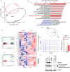
LXR-deficient mouse prostate reveals a decrease in mesenchymal transcriptomic signature. (A) HE-stained sections of dorsal prostates from WT mice and Lxrαβ-/- mice. Scale bars: upper panel: 500 µm; lower panel: 100 µm (B) Principal component analysis from RNAseq datasets of prostate WT mice and LXRαβ-/- mice capturing 55% (dimension 1) and 19% (dimension 2) of the data variability. (C) GSEA hallmark enrichment scores were ranked comparing signatures from WT and Lxrαβ-/- mouse prostates. (D) GSEA analysis plots for epithelial–mesenchymal transition and cholesterol homeostasis gene clusters and corresponding leading-edge gene set heatmap for each of them (NES: normalized enrichment score, FDR: false discovery rate).
Figure 4
Figure 4
LXR activation by GW3965 enhances the migration and invasion properties of PC3 cells. (A) Invasion assays were performed using Boyden chambers. PC3, DU145, and LNCaP cells seeded in culture medium without FBS were placed in the upper chamber previously coated with Matrigel. Medium with FBS was added in the lower chamber and the cells were treated with GW3965 (0.1 or 1 µM) for 24 h for each cell line. (B) Representative pictures of invaded cells. (C) Wound healing assays were performed to determine the effect of LXR activation on migration capacities. PC3 cells were treated with GW3965 (0.1 or 1 µM) and analyzed following 6 h, 12 h, 18 h, and 24 h of treatment. For each time point, relative wound closure was evaluated using Incucyte station. (D) Representative pictures of migrated cells. Scale bars: 400 µm. Data are expressed as the means ± SD. Mann–Whitney test statistical analysis; * p < 0.05, *** p < 0.001.
Figure 5
Figure 5
GW3965 treatment stimulates Vimentin accumulation in PC3 cells. (A) Representative Western blots of Fibronectin, Vimentin, N-Cadherin, and E-Cadherin accumulation using PC3 cells treated with DMSO (control) and GW3965 (0.1 or 1 µM). VIM accumulation is significantly increased in PC3 cells treated with 1 µM of GW3965. GAPDH was used as a loading control. (B) Immunofluorescence of PC3 cells stained for VIM (Fire scale). Scale bar: 100 µM. (C) ABCA1, SREBF1 (LXR target genes), and FN, VIM, CDH1, and CDH2 (mesenchymal genes) expressions were normalized using GAPDH in cells treated with DMSO or GW3965 (0.1 and 1 µM). (n = 6 per condition). Data are expressed as the means ± SD. Mann–Whitney test statistical analysis; * p < 0.05, ** p < 0.01. Original western blots are presented in File S3.
Figure 6
Figure 6
Molecular signature of GW3965 treatment reveals AREG as a potential target gene to control epithelial to mesenchymal transition. (A) Principal component analysis of PC3 cells treated with DMSO vs. GW3965 (1 µM) for 24 h, capturing 51% (dimension 1) and 23% (dimension 2) of the data variability. (B) GSEA hallmark rankings of enrichment scores in PC3 cells treated or not with GW3965 were plotted. (C) GSEA analysis plots for epithelial–mesenchymal transition and cholesterol homeostasis gene clusters and a corresponding leading-edge gene set heatmap for each of them. (D) Venn diagram summarizing the overlap between DRGs of PC3 cells treated with DMSO vs. GW3965 (left circle) and DRGs of prostates from WT vs. Lxrαβ-/- mice (right circle). This analysis leads to the identification of 28 genes deregulated in both datasets. (E) RNA-seq enrichment profiles for AREG locus coding sequences in PC3 cells treated with DMSO or with GW3965 (0.1 or 1 µM) for 24 h (n = 5 per condition) correlated with the RT-qPCR analysis of AREG expression using a similar experimental design (n = 6 per condition) (F) Representative Western blots of Amphiregulin accumulation from three independent experiments in PC3 cells treated with DMSO, GW3965 (1 µM), T0901317 (1 µM), or SR4593 (1 µM). Alternatively, PC3 cells treated with DMSO or GW3965 (1 µM) with cycloheximide (CHX) (50 µg/mL), an inhibitor of translation (n = 3 per condition). ABCG1 protein accumulation was monitored as a control of LXR activation, with ACTIN as a loading control. Three forms of Amphiregulin (AREG) were detected: Pro-Amphiregulin, Mature Amphiregulin, and the soluble form (Black arrows). Data are expressed as the means ± SD. Mann–Whitney test statistical analysis; ** p < 0.001. Original western blots are presented in File S3.
Figure 7
Figure 7
GW3965 increases Vimentin accumulation and the spindle-shape PC3 cell phenotype in a model of NSG orthotopically grafted mice. (A) Experimental procedure to study the metastatic spread of human advanced prostate cancer cells in NSG host mice. PC3-GFP were xenografted in NSG mice. One week after the surgery, mice were treated (intraperitoneal injection) with either DMSO or an LXR agonist (GW3965, 10 mg/kg), twice a week. Mice were sacrificed 5 weeks post-implantation, and prostates, lungs, lymph nodes, and kidneys were collected. (B) Representative pictures of implantation primary site (prostate) obtained during necropsy. GFP-fluorescence and HE-stained sections of the prostate confirm the graft success compared to non-grafted mice. (C) GFP immunodetection on the primary site of implantation observed using mice treated with DMSO or GW3965. Higher magnification exhibits the tumor border (filled arrows) compared to the normal tissue (open arrows) negative for the GFP. (D,E) Hematoxylin and eosin staining was used to analyze cellular spindle shape in vehicle- or GW3965-treated mice. Cell circularity was revealed with a red hot scale after segmentation and quantified using image J software. (F) Vimentin accumulation was detected (orange hot scale) in tumors from both vehicle- or GW3965-treated mice. Scale bar: 100 µm.
Figure 8
Figure 8
GW3965 increases PC3 invasiveness in NSG orthotopically grafted mice. (A) Representative pictures of the peritoneal cavity from vehicle- or GW3965-treated PC3-grafted mice. (B) Number of metastatic lymph nodes counted in the peritoneal cavity during necropsy. (C) Representative pictures of lungs obtained during necropsy. GFP-fluorescence exhibits an increase in fluorescent signals in mice treated with GW3965. (D,E) Lungs were analyzed by immunofluorescence detection of GFP (green) and Vimentin or Amphiregulin (red and fire scale), white arrowheads indicated metastasis with GFP and Amphiregulin positive staining. Scale bar: 100 µm. Data are expressed as the means ± SD. Mann–Whitney test statistical analysis; * p < 0.05.

References

    1. Sung H., Ferlay J., Siegel R.L., Laversanne M., Soerjomataram I., Jemal A., Bray F. Global Cancer Statistics 2020: GLOBOCAN Estimates of Incidence and Mortality Worldwide for 36 Cancers in 185 Countries. CA Cancer J. Clin. 2021;71:209–249. doi: 10.3322/caac.21660. - DOI - PubMed
    1. Brabletz T., Kalluri R., Nieto M.A., Weinberg R.A. EMT in Cancer. Nat. Rev. Cancer. 2018;18:128–134. doi: 10.1038/nrc.2017.118. - DOI - PubMed
    1. Mock K., Preca B.-T., Brummer T., Brabletz S., Stemmler M.P., Brabletz T. The EMT-Activator ZEB1 Induces Bone Metastasis Associated Genes Including BMP-Inhibitors. Oncotarget. 2015;6:14399–14412. doi: 10.18632/oncotarget.3882. - DOI - PMC - PubMed
    1. Ren J., Chen Y., Song H., Chen L., Wang R. Inhibition of ZEB1 Reverses EMT and Chemoresistance in Docetaxel-Resistant Human Lung Adenocarcinoma Cell Line. J. Cell Biochem. 2013;114:1395–1403. doi: 10.1002/jcb.24481. - DOI - PubMed
    1. Shibue T., Weinberg R.A. EMT, CSCs, and Drug Resistance: The Mechanistic Link and Clinical Implications. Nat. Rev. Clin. Oncol. 2017;14:611–629. doi: 10.1038/nrclinonc.2017.44. - DOI - PMC - PubMed

LinkOut - more resources