. 2024 Jul 24;11(8):746.
doi: 10.3390/bioengineering11080746.

PRF Lysates Modulate Chemokine Expression in Oral Squamous Carcinoma and Healthy Epithelial Cells

Affiliations

PRF Lysates Modulate Chemokine Expression in Oral Squamous Carcinoma and Healthy Epithelial Cells

Zohreh Afradi et al. Bioengineering (Basel). .

Abstract

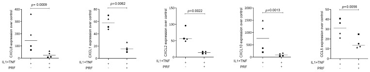
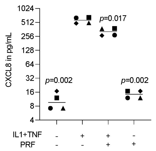
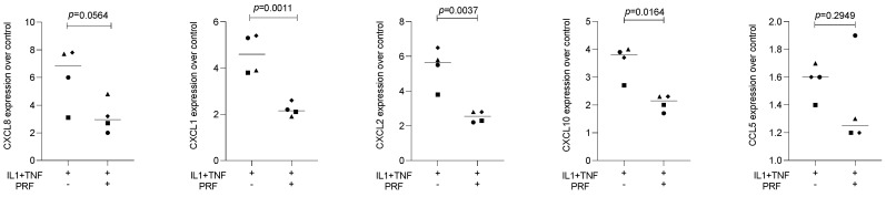
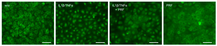
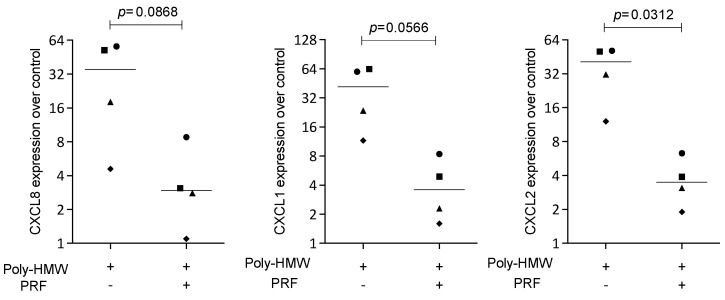
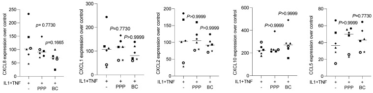
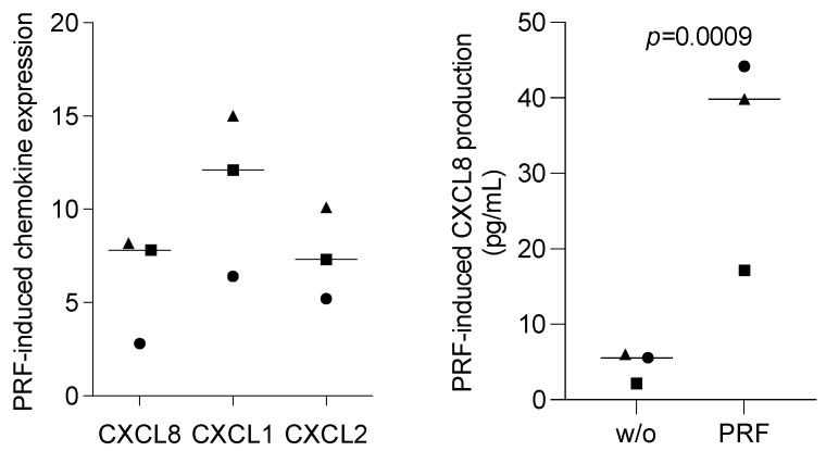
Platelet-rich fibrin (PRF), originally used to support soft tissue healing, is also considered a therapeutic option for treating oral lichen planus and leukoplakia. The progression from the two premalignant lesions to the aggressive malignant oral squamous cell carcinoma involves an inflammatory process linked to chemokine expression. Thus, there is a rationale for studying how PRF modulates the expression of chemokines in oral squamous carcinoma cells. To this aim, we expose the oral squamous carcinoma cell line HSC2 to IL1β and TNFα either alone or in the presence of lysates obtained from solid PRF membranes. We report here that in HSC2 cells, PRF lysates significantly reduce the forced transcription of chemokines, e.g., CXCL1, CXCL2, CXCL8, CXCL10, and CCL5. Moreover, PRF lysates attenuate the nuclear translocation of p65 in HSC2 oral epithelial cells when exposed to IL1β and TNFα. PRF lysates further reduce chemokine expression provoked by poly:IC HMW. Even though less pronounced, PRF lysates reduce IL1β- and TNFα-induced chemokine expression in TR146 cells. In primary oral epithelial cells, however, PRF lysates increase the basal expression of CXCL1, CXCL2 and CXCL8. Thus, PRF can exert a biphasic effect on chemokine expression in oral squamous cell carcinoma cell lines and primary oral epithelial cells. These findings suggest that PRF may reduce inflammation in a malignant environment while provoking an immunological response in healthy oral epithelium.

Keywords: HSC2; PRF; TR146; chemokines; inflammation; oral epithelial cells; oral squamous carcinoma cells; platelet-rich fibrin.


Conflict of interest statement

Salman Abbas Zadeh is an employee of epitome GmbH. Other authors declare no conflict of interest.

Figures

Figure 1
Figure 1
PRF lysates suppress IL1β- and TNFα-induced chemokine expression in HSC2 cells. The oral squamous cell carcinoma cell line HSC2 was exposed to IL1β and TNFα in the presence or absence of PRF lysates. Gene expression analysis was performed the following day. Data points represent four independent experiments. Data were normalized to untreated control cells with x-fold changes compared to the untreated cells. The statistical analysis was based on the ratio paired t-test and p-values reported.
Figure 2
Figure 2
PRF lysates suppress IL1β- and TNFα-induced CXCL8 protein in HSC2 cells. The oral squamous cell carcinoma cell line HSC2 was exposed to IL1β and TNFα in the presence or absence of PRF lysates. A CXCL8 immunoassay from HSC2 cell lysates was performed the following day. Data points represent four independent experiments. Data show CXCL8 in pg/mL of the cell lysates. The statistical analysis was based on the ratio paired t-test and p-values reported.
Figure 3
Figure 3
PRF lysates suppress IL1β- and TNFα-induced chemokine expression in TR146 cells. The oral squamous cell carcinoma cell line TR146 was exposed to IL1β and TNFα in the presence or absence of PRF lysates. Gene expression analysis was performed the following day. Data points represent four independent experiments. Data were normalized to untreated control cells with x-fold changes compared to the untreated cells. The statistical analysis was based on the ratio paired t-test and p-values reported.
Figure 4
Figure 4
PRF lysates reduced IL1β- and TNFα-induced p65 translocation in HSC2 cells. The oral squamous cell carcinoma cell line HSC2 was preexposed to 30% PRF lysates overnight before IL1β and TNFα were added for 45 min. Immunostaining indicated that PRF lysates reduced IL1β- and TNFα-induced p65 translocation in HSC2 cells. The scale bars represent 100 µm.
Figure 5
Figure 5
PRF lysates suppress IL1β- and TNFα-induced chemokine expression in HSC2 cells. The oral squamous cell carcinoma cell line HSC2 was exposed to poly(I:C) HMW in the presence or absence of PRF lysates. Gene expression analysis was performed the following day. Data points represent four independent experiments. Data were normalized to untreated control cells with x-fold changes compared to the untreated cells. The statistical analysis was based on the ratio paired t-test and p-values reported.
Figure 6
Figure 6
PPP and buffy coat lysates had no impact on IL1β- and TNFα-induced chemokine expression in HSC2 cells. The oral squamous cell carcinoma cell line HSC2 was exposed to IL1β and TNFα in the presence or absence of PPP and buffy coat (BC) lysates. Gene expression analysis was performed the following day. Data points represent independent experiments. Data were normalized to untreated control cells with x-fold changes compared to the untreated cells. The statistical analysis was based on the Friedmann test, and p-values were reported.
Figure 7
Figure 7
PRF lysates enhanced the CXCL8 production of primary oral epithelial cells. Primary oral epithelial cells were exposed to PRF lysates. Gene expression analysis was performed the following day. Data were normalized to untreated control cells with x-fold changes compared to the untreated cells (left panel). The supernatant was harvested and subjected to immunoassay (right panel). Data points represent three independent experiments. The statistical analysis was based on the ratio paired t-test and p-values.

Similar articles

References

    1. Moutsopoulos N.M., Konkel J.E. Tissue-Specific Immunity at the Oral Mucosal Barrier. Trends Immunol. 2018;39:276–287. doi: 10.1016/j.it.2017.08.005. - DOI - PMC - PubMed
    1. Williams D.W., Greenwell-Wild T., Brenchley L., Dutzan N., Overmiller A., Sawaya A.P., Webb S., Martin D., Genomics N.N., Computational Biology C., et al. Human oral mucosa cell atlas reveals a stromal-neutrophil axis regulating tissue immunity. Cell. 2021;184:4090–4104.e4015. doi: 10.1016/j.cell.2021.05.013. - DOI - PMC - PubMed
    1. Yuan X., Chen J., Van Brunt L.A., Grauer J., Xu Q., Pei X., Wang L., Zhao Y., Helms J.A. Formation and regeneration of a Wnt-responsive junctional epithelium. J. Clin. Periodontol. 2020;47:1476–1484. doi: 10.1111/jcpe.13371. - DOI - PMC - PubMed
    1. Gonzalez-Arriagada W.A., Canedo-Marroquin G., Adorno-Farias D., Fernandez-Ramires R. New insights into the role of the oral leukoplakia microenvironment in malignant transformation. Front. Oral Health. 2024;5:1363052. doi: 10.3389/froh.2024.1363052. - DOI - PMC - PubMed
    1. Chiang C.P., Yu-Fong Chang J., Wang Y.P., Wu Y.H., Lu S.Y., Sun A. Oral lichen planus—Differential diagnoses, serum autoantibodies, hematinic deficiencies, and management. J. Formos. Med. Assoc. 2018;117:756–765. doi: 10.1016/j.jfma.2018.01.021. - DOI - PubMed