. 2024 Aug 13;14(16):2336.
doi: 10.3390/ani14162336.

Immunolocalization of Two Neurotrophins, NGF and BDNF, in the Pancreas of the South American Sea Lion Otaria flavescens and Bottlenose Dolphin Tursiops truncatus

Affiliations

Immunolocalization of Two Neurotrophins, NGF and BDNF, in the Pancreas of the South American Sea Lion Otaria flavescens and Bottlenose Dolphin Tursiops truncatus

Claudia Gatta et al. Animals (Basel). .

Abstract

In this study, we have investigated the immunolocalization of NGF (Nerve Growth Factor) and BDNF (Brain-Derived Neurotrophic Factor) in the pancreas of two species of marine mammals: Tursiops truncatus (common bottlenose dolphin), belonging to the order of the Artiodactyla, and Otaria flavescens (South American sea lion), belonging to the order of the Carnivora. Our results demonstrated a significant presence of NGF and BDNF in the pancreas of both species with a wide distribution pattern observed in the exocrine and endocrine components. We identified some differences that can be attributed to the different feeding habits of the two species, which possess a different morphological organization of the digestive system. Altogether, these preliminary observations open new perspectives on the function of neurotrophins and the adaptive mechanisms of marine mammals in the aquatic environment, suggesting potential parallels between the physiology of marine and terrestrial mammals.

Keywords: Brain-Derived Neurotrophic Factor; Nerve Growth Factor; South American sea lion; common bottlenose dolphin; marine mammals; pancreas.


Conflict of interest statement

The authors declare no conflicts of interest.

Figures

Figure 1
Figure 1
Hematoxylin eosin staining of the pancreas of Otaria flavescens and Tursiops truncatus. (AD) Otaria flavescens. Cells in the parenchyma and large elongate islet (arrow) (A); Langerhans islets of medium size and ovoid shape (arrow) and blood vessels (arrowhead) (B); blood vessels in the interconnective septa (arrowhead) (C); ganglion with neurons in the parenchyma (dotted arrow) (D). (EH) Tursiops truncatus. Exocrine cells in the parenchyma and islet of Langerhans of small size with ovoid shape (arrow) (E); islets of Langerhans of medium size (arrow) (F); islet of Langerhans small size (arrow) and blood vessels (arrowhead) (G); blood vessels (arrowhead) (H). Scale bar 100 µm.
Figure 2
Figure 2
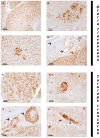
NGF immunoreactivity in the pancreas of Otaria flavescens and Tursiops truncatus. (AD) Otaria flavescens. Positive exocrine cells (A); positive islets of Langerhans (B); positive fibers in the vessel wall (arrow) in the interconnective septa (C); ganglion with positive neurons (asterisk) and fibers (arrowhead) (D). (EH) Tursiops truncatus. Positive exocrine cells in parenchyma (E); small islets of Langerhans (F); immunoreactivity in fibers along blood vessels wall (arrow) (G); positive fibers bundle (arrowhead) (H). Scale bar 100 µm.
Figure 3
Figure 3
BDNF immunoreactivity in the pancreas of Otaria flavescens and Tursiops truncatus. (AD) Otaria flavescens. BDNF immunorectivity of exocrine cells in parechima (A); positive large islet of Langerhans (B); positive medium islet of Langerhans (C); positive fibers in the wall of blood vessels (arrow), positive neurons (asterisk) and fibers (arrowhead) in the parenchyma (D); (EH) Tursiops truncatus. Immunoreactivity of exocrine cells in parenchyma (E); positive islet of Langerhans (F); positive fibers in the wall of blood vessels (arrow) (G); immunoreactive fibers (arrowhead for fiber bundle) (H). Scale bar 100 µm.

Similar articles

Cited by

References

    1. Liu S. Neurotrophic factors in enteric physiology and pathophysiology. Neurogastroenterol. Motil. 2018;30:e13446. doi: 10.1111/nmo.13446. - DOI - PMC - PubMed
    1. Gotz R., Koster R., Winkler C., Raulft F., Lottspeich F., Schartl M., Thoenen H. Neurotrophin-6 is a new member of the nerve growth factor family. Nature. 1994;372:266–269. doi: 10.1038/372266a0. - DOI - PubMed
    1. Lai K.O., Fu W.Y., Ip F.C., Ip N.Y. Cloning and expression of a novel neurotrophin, NT-7, from Carp. Mol. Cell. Neurosci. 1998;11:64–76. doi: 10.1006/mcne.1998.0666. - DOI - PubMed
    1. Nilsson A.S., Fainzilber M., Falck P., Ibañez C.F. Neurotrophin-7: A novel member of the neurotrophin family from the zebrafish. FEBS Lett. 1998;424:285–290. doi: 10.1016/S0014-5793(98)00192-6. - DOI - PubMed
    1. Kaplan D.R., Martin-Zanca D., Parada L.F. Tyrosine phosphorylation and tyrosine kinase activity of the trk protooncogene product induced by NGF. Nature. 1991;350:158–160. doi: 10.1038/350158a0. - DOI - PubMed

LinkOut - more resources