. 2024 Jul 26;13(8):701.
doi: 10.3390/antibiotics13080701.

The pH-Insensitive Antimicrobial and Antibiofilm Activities of the Frog Skin Derived Peptide Esc(1-21): Promising Features for Novel Anti-Infective Drugs

Affiliations

The pH-Insensitive Antimicrobial and Antibiofilm Activities of the Frog Skin Derived Peptide Esc(1-21): Promising Features for Novel Anti-Infective Drugs

Maria Rosa Loffredo et al. Antibiotics (Basel). .

Abstract

The number of antibiotic-resistant microbial infections is dramatically increasing, while the discovery of new antibiotics is significantly declining. Furthermore, the activity of antibiotics is negatively influenced by the ability of bacteria to form sessile communities, called biofilms, and by the microenvironment of the infection, characterized by an acidic pH, especially in the lungs of patients suffering from cystic fibrosis (CF). Antimicrobial peptides represent interesting alternatives to conventional antibiotics, and with expanding properties. Here, we explored the effects of an acidic pH on the antimicrobial and antibiofilm activities of the AMP Esc(1-21) and we found that it slightly lost activity (from 2- to 4-fold) against the planktonic form of a panel of Gram-negative bacteria, with respect to a ≥ 32-fold of traditional antibiotics. Furthermore, it retained its activity against the sessile form of these bacteria grown in media with a neutral pH, and showed similar or higher effectiveness against the biofilm form of bacteria grown in acidic media, simulating a CF-like acidic microenvironment, compared to physiological conditions.

Keywords: Gram-negative bacteria; acidic pH; antibiotics; antimicrobial peptides; biofilm; cystic fibrosis; infections.


Conflict of interest statement

The authors declare no conflicts of interest.

Figures

Figure 1
Figure 1
Variation in the MIC (expressed as fold-change in MIC value) of AMPs and antibiotics against A. baumannii ATCC 19606, E. coli ATCC 25922 and P. aeruginosa ATCC 27853, in 10% TSB at different pH values. The plotted data represent the modal values of three independent experiments.
Figure 2
Figure 2
Variation in the MIC values of Esc(1-21) and tobramycin at different pH values against several Gram-negative strains, including reference and multidrug-resistant clinical isolates, in 10% TSB at different pH values. The data are the modal values from three independent experiments.
Figure 3
Figure 3
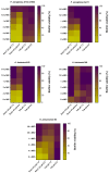
The effect of Esc(1-21) and tobramycin (Tob), against 20 h-preformed biofilms of several Gram-negative strains in LB and treated with the peptide/antibiotic for 2 h in PBS at two different pHs, i.e., 7.5 and 6.5. The colors represent the percentage of biofilm viability from 0% (yellow) to 100% (purple). Data were collected from three independent experiments.
Figure 4
Figure 4
Percentage of biofilm viability of different Gram-negative strains in m63 at pH 6.5 with respect to pH 7.5 (set as 100%). The colors represent the percentage of biofilm viability from 0% (light green) to 100% (dark green). Data were collected from three independent experiments. The statistical analysis was conducted with the t test for all strains between the two different pH values: all differences were found to be significant (p < 0.05), with the sole exception of A. baumannii #4 (p = 0.765).
Figure 5
Figure 5
The effect of Esc(1-21) and tobramycin (Tob), against preformed biofilms of several Gram-negative bacterial strains in m63 medium at two different pHs, i.e., 7.5 and 6.5. Data were collected from three independent experiments.
Figure 6
Figure 6
Kinetics of the effect of different concentrations of Esc(1-21) at pH 7.5 and 6.5, on the leakage of CyF encapsulated into POPE/POPG LUVs. LUVs were used at a final lipid concentration of 100 µM. Data points are the mean of three different experiments. SD is not shown for clarity.
Figure 7
Figure 7
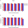
Viability of HaCaT (A) and A549 (B) cells after 24 h treatment with different concentrations of Esc(1-21) in DMEMg at pH 7.5 and 6.5. Cells not treated with peptides were used as controls. All data are the means of three replicates ± standard error of the mean (SEM). The statistical analysis was conducted with the t test for all concentrations between the two different pH values; all differences were found to be not significant (p > 0.05), with the only exception of 140 μg/mL against A549 cells (pH 7.5 vs. pH 6.5, p < 0.05).

References

    1. McDonald E.F., Meiler J., Plate L. CFTR Folding: From Structure and Proteostasis to Cystic Fibrosis Personalized Medicine. ACS Chem. Biol. 2023;18:2128–2143. doi: 10.1021/acschembio.3c00310. - DOI - PMC - PubMed
    1. Brown S.D., White R., Tobin P. Keep them breathing: Cystic fibrosis pathophysiology, diagnosis, and treatment. JAAPA. 2017;30:23–27. doi: 10.1097/01.JAA.0000515540.36581.92. - DOI - PubMed
    1. Pezzulo A.A., Tang X.X., Hoegger M.J., Abou Alaiwa M.H., Ramachandran S., Moninger T.O., Karp P.H., Wohlford-Lenane C.L., Haagsman H.P., van Eijk M., et al. Reduced airway surface pH impairs bacterial killing in the porcine cystic fibrosis lung. Nature. 2012;487:109–113. doi: 10.1038/nature11130. - DOI - PMC - PubMed
    1. Simonin J., Bille E., Crambert G., Noel S., Dreano E., Edwards A., Hatton A., Pranke I., Villeret B., Cottart C.H., et al. Airway surface liquid acidification initiates host defense abnormalities in Cystic Fibrosis. Sci. Rep. 2019;9:6516. doi: 10.1038/s41598-019-42751-4. - DOI - PMC - PubMed
    1. McShane D., Davies J.C., Davies M.G., Bush A., Geddes D.M., Alton E.W. Airway surface pH in subjects with cystic fibrosis. Eur. Respir. J. 2003;21:37–42. doi: 10.1183/09031936.03.00027603. - DOI - PubMed

LinkOut - more resources