Review
. 2024 Aug 22;13(8):788.
doi: 10.3390/antibiotics13080788.

Artificial Intelligence-Driven Analysis of Antimicrobial-Resistant and Biofilm-Forming Pathogens on Biotic and Abiotic Surfaces

Affiliations
Review

Artificial Intelligence-Driven Analysis of Antimicrobial-Resistant and Biofilm-Forming Pathogens on Biotic and Abiotic Surfaces

Akanksha Mishra et al. Antibiotics (Basel). .

Abstract

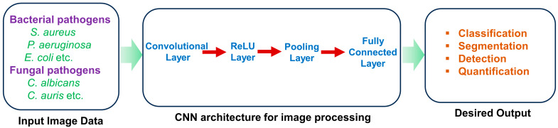
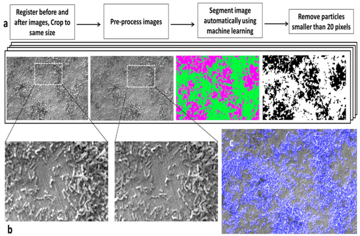
The growing threat of antimicrobial-resistant (AMR) pathogens to human health worldwide emphasizes the need for more effective infection control strategies. Bacterial and fungal biofilms pose a major challenge in treating AMR pathogen infections. Biofilms are formed by pathogenic microbes encased in extracellular polymeric substances to confer protection from antimicrobials and the host immune system. Biofilms also promote the growth of antibiotic-resistant mutants and latent persister cells and thus complicate therapeutic approaches. Biofilms are ubiquitous and cause serious health risks due to their ability to colonize various surfaces, including human tissues, medical devices, and food-processing equipment. Detection and characterization of biofilms are crucial for prompt intervention and infection control. To this end, traditional approaches are often effective, yet they fail to identify the microbial species inside biofilms. Recent advances in artificial intelligence (AI) have provided new avenues to improve biofilm identification. Machine-learning algorithms and image-processing techniques have shown promise for the accurate and efficient detection of biofilm-forming microorganisms on biotic and abiotic surfaces. These advancements have the potential to transform biofilm research and clinical practice by allowing faster diagnosis and more tailored therapy. This comprehensive review focuses on the application of AI techniques for the identification of biofilm-forming pathogens in various industries, including healthcare, food safety, and agriculture. The review discusses the existing approaches, challenges, and potential applications of AI in biofilm research, with a particular focus on the role of AI in improving diagnostic capacities and guiding preventative actions. The synthesis of the current knowledge and future directions, as described in this review, will guide future research and development efforts in combating biofilm-associated infections.

Keywords: abiotic surfaces; antimicrobial resistance; artificial intelligence; biofilm formation; biotic surfaces; deep learning; image processing; machine learning; microbial pathogens.


Conflict of interest statement

The authors declare no conflicts of interest.

Figures

Figure 8
Figure 8
Confocal laser scanning microscopy allowed nanosensor fluorescence imaging in reference buffers. (a) FITC (green) and NR (red) nanosensor fluorescence were observed in 8 reference buffers. Z-stack imaging captured biofilm depth at each pH. Representative 2D pictures with overlays improve clarity. A 100 µm scale bar is used for all pictures. (b) FITC/NR fluorescence intensity (FI) was plotted versus pH. Error bars represent standard deviation, while blue-shaded regions represent fitted curve confidence intervals. Reprinted from [146]. Copyright © 2022 by the authors and Licensee, Scientific Reports.
Figure 1
Figure 1
The biofilms created by bacterial pathogens are present on various human body tissues, medical equipment, such as catheters or prostheses, and in the environment. It acts as a reservoir for potential infections that may occur in the future. The common bacterial species that are associated with diseases caused by biofilms are depicted in a schematic figure on the left. The arrows in the diagram indicate where these bacteria are located throughout the body. When it comes to the development of biofilm, attachment, maturation, and separation are all essential components (bottom right). Multiple components contribute to this multistep process, including bacterial surface chemicals, secreted matrix effectors, ambient components, and stressors. Therefore, the regulation of bacterial biofilm (located on the lower right) necessitates the utilization of intricate positive and negative regulatory mechanisms. Second messengers such as Bis-(3′-5′)-cyclic dimeric guanosine monophosphate (c-di-GMP) are examples of these mechanisms. Quorum sensing (QS), regulatory sRNAs, alternative sigma factors, two-component systems, and other mechanisms are also included. Reprinted from [26], Copyright © 2021 by the authors and Shared Science Publishers OG. Enterohemorrhagic Escherichia coli (EHEC), extracellular DNA (eDNA), and small RNAs (sRNAs).
Figure 2
Figure 2
Global annual economic burden of various market sectors with biofilm-associated technologies (all values are in billions of dollars). The information obtained from [68].
Figure 3
Figure 3
Global annual economic burden of various biofilm-associated infections on the health sector (all values are in billions of dollars). The information obtained from [68].
Figure 4
Figure 4
Basic block diagram of machine learning and deep learning. Reprinted from [105]. Copyright © 2021 by the authors and Licensee, J Big Data.
Figure 5
Figure 5
Internal workflow diagram of deep learning. Reprinted from the [105] Copyright © 2021 by the authors and Licensee, J Big Data.
Figure 6
Figure 6
Deep learning architecture represents the detection of microorganisms from microscopic images.
Figure 7
Figure 7
(a) A method was devised to segment biofilm from surfaces in SEM images. (b) Close-up images illustrate the surface before and after pre-processing, where outliers were eliminated to smooth scratches on the surface, facilitating improved segmentation. (c) An overlay demonstrates the segmented area (highlighted in blue) overlaid onto the SEM image, showcasing precise biofilm detection. Reprinted from [121]. Copyright © 2016 by the authors and Licensee, Scientific Reports.

Similar articles

Cited by

References

    1. Nabadda S., Kakooza F., Kiggundu R., Walwema R., Bazira J., Mayito J., Mugerwa I., Sekamatte M., Kambugu A., Lamorde M. Implementation of the World Health Organization global antimicrobial resistance surveillance system in Uganda, 2015–2020: Mixed-methods study using national surveillance data. JMIR Public Health Surveill. 2021;7:e29954. doi: 10.2196/29954. - DOI - PMC - PubMed
    1. Centers for Disease Control and Prevention . Antibiotic Resistance Threats in the United States, 2019. US Department of Health and Human Services, Centres for Disease Control and Prevention; Atlanta, GA, USA: 2019.
    1. ECDC. EMEA . The Bacterial Challenge–Time to React a Call to Narrow the Gap between Multidrug-Resistant Bacteria in the EU and Development of New Antibacterial Agents. ECDC & EMEA; Solna, Sweden: 2009.
    1. Ventola C.L. The antibiotic resistance crisis: Part 1: Causes and threats. Pharm. Ther. 2015;40:277–283. - PMC - PubMed
    1. Davies J., Davies D. Origins and evolution of antibiotic resistance. Microbiol. Mol. Biol. Rev. 2010;74:417–433. doi: 10.1128/MMBR.00016-10. - DOI - PMC - PubMed

LinkOut - more resources