. 2024 Jul 23;12(8):1634.
doi: 10.3390/biomedicines12081634.

The Aggregation of α-Synuclein in the Dorsomedial Striatum Significantly Impairs Cognitive Flexibility in Parkinson's Disease Mice

Affiliations

The Aggregation of α-Synuclein in the Dorsomedial Striatum Significantly Impairs Cognitive Flexibility in Parkinson's Disease Mice

Jing Chen et al. Biomedicines. .

Abstract

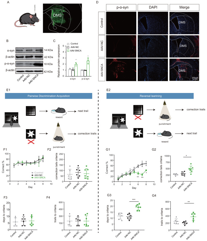
This study focused on α-synuclein (α-syn) aggregation in the dorsomedial striatum (DMS) so as to investigate its role in the cognitive flexibility of Parkinson's disease (PD). Here, we investigated the cognitive flexibility by assessing reversal learning abilities in MPTP-induced subacute PD model mice and in C57BL/6J mice with α-syn aggregation in the DMS induced by adenovirus (AAV-SNCA) injection, followed by an analysis of the target protein's expression and distribution. PD mice exhibited impairments in reversal learning, positively correlated with the expression of phosphorylated α-syn in the DMS. Furthermore, the mice in the AAV-SNCA group exhibited reversal learning deficits and a reduction in acetylcholine levels, accompanied by protein alterations within the DMS. Notably, the administration of a muscarinic receptor 1 (M1R) agonist was able to alleviate the aforementioned phenomenon. These findings suggest that the impaired cognitive flexibility in PD may be attributed to the diminished activation of acetylcholine to M1R caused by α-syn aggregation.

Keywords: Parkinson’s disease; acetylcholine; cognitive flexibility; muscarinic receptor 1; reversal learning; α-synuclein.


Conflict of interest statement

The authors declare no conflicts of interest.

Figures

Figure 1
Figure 1
Mode diagram of small animal touch screen system.
Figure 2
Figure 2
MPTP subacute Parkinson’s disease (PD) model was established. (A) The timeline for experimental arrangement. (B1,B2) Rotarod test and (C1,C2) Open-field test results suggested that the mice of MPTP group exhibited motor disability (n = 15–16 mice, ** p < 0.01, *** p < 0.001 vs. control group). (D1,D2) Western Blotting (WB) results for TH in substantia nigra, showed decreased expression in MPTP group (n = 3, *** p < 0.001 vs. control group). (E1,E2) Immunofluorescence (IF) results showed that the distribution of TH+ positive neurons decreased in MPTP group (n = 5, *** p < 0.001 vs. control group). Scale bar: 200 µm.
Figure 3
Figure 3
The PD mice demonstrate impaired reversal learning abilities associated with the accumulation of alpha-synuclein (α-syn). (A) Behavioral paradigm diagram of the modified Morris water maze (MWM). (B) The latency target index (referring to the time taken by the mice to reach the plateau quadrant for the first time during testing) serves as an indicator of reversal learning performance; low index indicates poor reversal learning in the MPTP group ((training test − reversal test)/(training test + reversal test); n = 15–16 mice, ** p < 0.01 vs. control group). The lower the index, the worse the ability in reversal learning. (C) Time spent in each quadrant by mice in the reversal learning phase showed that the MPTP group spent relatively more time in the original quadrant compared to the control group (n = 15–16 mice). (D,E) WB results for α-syn and p-α-syn in the dorsomedial striatum, showed increased expression in the low-index (index ≤ 0.85) group (n = 3; * p < 0.05, ** p < 0.01 vs. control group; # p < 0.05 vs. high group (index > 0.85)); while, the statistical difference in the levels of α-syn between the Index high and Index low groups was not significant. (F) Pearson correlation analysis of p-α-syn expression and reverse learning index (n = 17 mice, r = −0.932, p < 0.01).
Figure 4
Figure 4
Reversal learning deficits were observed upon accumulation of α-syn in the dorsomedial striatum. (A) Schematic diagram of cerebral stereotactic drug delivery, and AAV-SNCA spread region in the striatum visualized in green. (B,C) WB results for α-syn and p-α-syn in the striatum showing increased expression in the AAV-SNCA group (n = 3, ** p < 0.01 vs. AAV-NC group). (D) IF results for striatal p-α-syn (scale bar: 200 μm). (E1,E2) Behavioral paradigm diagram of pairwise discrimination acquisition phases and reversal learning phases in the small animal touchscreen systems. (F1) Correctness per day for each group in the pairwise discrimination learning phase when 50% of the mice reach the criterion (>80% correct on two consecutive days). (F2F4) The number of errors corrected, learning sessions and time required to achieve the criterion in the pairwise discrimination learning phase. (G1) Correctness per day for each group in the reversal phase when 50% of the mice reach the criterion. (G2G4) The number of errors corrected, learning sessions and time required to achieve the criterion in the reversal phase. (n = 6–7 mice, * p < 0.05, ** p < 0.01, *** p < 0.001 vs. AAV-NC group.
Figure 5
Figure 5
The aggregation of α-syn in the dorsomedial striatum inhibited acetylcholine (Ach) release from cholinergic interneurons (ChI). (AC) WB results and statistical analysis of NR2D and Choline acetyl transferase (ChAT) in the dorsomedial striatum (n = 3, ** p < 0.01 vs. AAV-NC group); however, there was no statistically significant difference observed in the levels of ChAT between the AAV-NC group and the AAV-SNCA group. (D) ELISA results showed a decrease in ACh content in the dorsomedial striatum of mice in the AAV-SNCA group (n = 8, ** p < 0.01 vs. AAV-NC group). (EG) WB results and statistical analysis of Snap25 and Syntaxin in the dorsomedial striatum (n = 3, ** p < 0.01 vs. AAV-NC group).
Figure 6
Figure 6
The aggregation of α-syn led to diminished activation on indirect pathway spiny projection neurons (iSPNs). (AC) WB results and statistical analysis of c-Fos and M1R in the dorsomedial striatum. (D,E) The WB results were obtained for phosphorylated Erk (p-Erk), total Erk, and the ratio of p-Erk to Erk expression. (F,G) The WB results were obtained for phosphorylated AKT (p-AKT), total AKT, and the ratio of p-AKT to AKT expression. (HJ) WB results and statistical analysis of PSD 95 and CaMKII in the dorsomedial striatum. (n = 3, * p < 0.05 vs. AAV-NC group).
Figure 7
Figure 7
M1R activation mitigates the reversal learning deficits induced by α-syn aggregation in mice. (A) Experimental flow chart of drug administration and small animal touchscreen systems. (B,C) Statistical analysis of the time required and the number of errors corrected to achieve the criterion in the pairwise discrimination learning phase (n = 12 mice). (D,E) Statistical analysis of the time required and the number of errors corrected to achieve the criterion in the first reversal learning phase (n = 12 mice, ** p < 0.01 vs. AAV-NC group). (F,G) Statistical analysis of the time required and the number of errors corrected to achieve the criterion in the second reversal learning phase (n = 6 mice, * p < 0.05, ** p < 0.01 vs. AAV-SNCA + DMSO group); however, there was no significant difference between AAV-NC + DMSO and AAV-NC + VU0357017 groups. (H,I) Before and after intraperitoneal injection of M1R activator, statistical analysis of the time required and the number of errors corrected to achieve the criterion in the second reversal learning phase. A significant reduction in both the time required to achieve the criterion and the number of error corrections in the mice of the AAV-SNCA group were observed after intraperitoneal injection of U0357017 (** p < 0.01), whereas such effects were conspicuously absent in the AAV-NC group. Additionally, no significant differences were observed in these parameters in the two groups (AAV-NC and AAV-SNCA groups) before and after intraperitoneal injection of DMSO.
Figure 8
Figure 8
Summary diagram.

Similar articles

References

    1. Ahmad M.H., Rizvi M.A., Ali M., Mondal A.C. Neurobiology of depression in Parkinson’s disease: Insights into epidemiology, molecular mechanisms and treatment strategies. Ageing Res. Rev. 2023;85:101840. doi: 10.1016/j.arr.2022.101840. - DOI - PubMed
    1. Santos-Garcia D., de Deus Fonticoba T., Cores Bartolome C., Feal Painceiras M.J., Iniguez-Alvarado M.C., Garcia Diaz I., Jesus S., Buongiorno M.T., Planellas L., Cosgaya M., et al. Staging Parkinson’s Disease According to the MNCD (Motor/Non-motor/Cognition/Dependency) Classification Correlates with Disease Severity and Quality of Life. J. Park. Dis. 2023;13:379–402. doi: 10.3233/JPD-225073. - DOI - PMC - PubMed
    1. Wang Z., Flores I., Donahue E.K., Lundquist A.J., Guo Y., Petzinger G.M., Jakowec M.W., Holschneider D.P. Cognitive flexibility deficits in rats with dorsomedial striatal 6-hydroxydopamine lesions tested using a three-choice serial reaction time task with reversal learning. Neuroreport. 2020;31:1055–1064. doi: 10.1097/WNR.0000000000001509. - DOI - PubMed
    1. Trenado C., Boschheidgen M., Rubenach J., N’Diaye K., Schnitzler A., Mallet L., Wojtecki L. Assessment of Metacognition and Reversal Learning in Parkinson’s Disease: Preliminary Results. Front. Hum. Neurosci. 2018;12:343. doi: 10.3389/fnhum.2018.00343. - DOI - PMC - PubMed
    1. Castane A., Theobald D.E., Robbins T.W. Selective lesions of the dorsomedial striatum impair serial spatial reversal learning in rats. Behav. Brain Res. 2010;210:74–83. doi: 10.1016/j.bbr.2010.02.017. - DOI - PMC - PubMed

LinkOut - more resources