. 2024 Jul 25;12(8):1665.
doi: 10.3390/biomedicines12081665.

Leveraging Hypotension Prediction Index to Forecast LPS-Induced Acute Lung Injury and Inflammation in a Porcine Model: Exploring the Role of Hypoxia-Inducible Factor in Circulatory Shock

Affiliations

Leveraging Hypotension Prediction Index to Forecast LPS-Induced Acute Lung Injury and Inflammation in a Porcine Model: Exploring the Role of Hypoxia-Inducible Factor in Circulatory Shock

Yuan-Ming Tsai et al. Biomedicines. .

Abstract

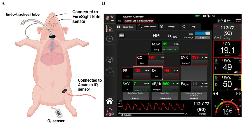
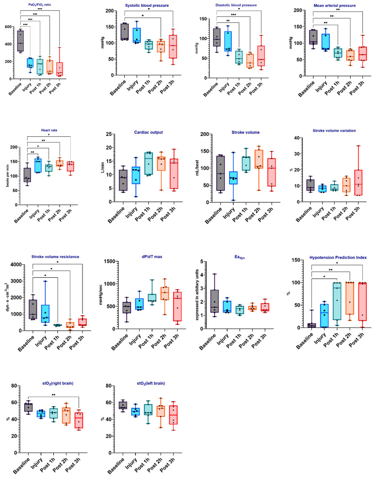
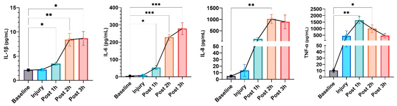
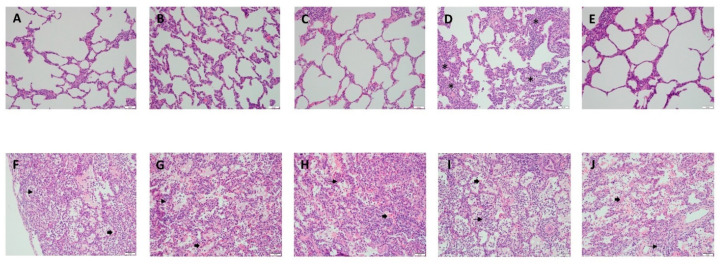
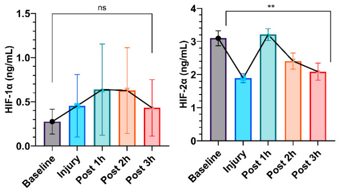
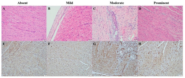
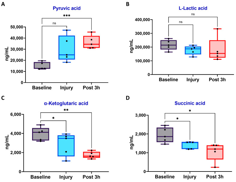
Acute respiratory distress syndrome (ARDS) is a critical illness in critically unwell patients, characterized by refractory hypoxemia and shock. This study evaluates an early detection tool and investigates the relationship between hypoxia and circulatory shock in ARDS, to improve diagnostic precision and therapy customization. We used a porcine model, inducing ARDS with mechanical ventilation and intratracheal plus intravenous lipopolysaccharide (LPS) injection. Hemodynamic changes were monitored using an Acumen IQ sensor and a ForeSight Elite sensor connected to the HemoSphere platform. We evaluated tissue damage, inflammatory response, and hypoxia-inducible factor (HIF) alterations using enzyme-linked immunosorbent assay and immunohistochemistry. The results showed severe hypotension and increased heart rates post-LPS exposure, with a notable rise in the hypotension prediction index (HPI) during acute lung injury (p = 0.024). Tissue oxygen saturation dropped considerably in the right brain region. Interestingly, post-injury HIF-2α levels were lower at the end of the experiment. Our findings imply that the HPI can effectively predict ARDS-related hypotension. HIF expression levels may serve as possible markers of rapid ARDS progression. Further research should be conducted on the clinical value of this novel approach in critical care, as well as the relationship between the HIF pathway and ARDS-associated hypotension.

Keywords: hemodynamic monitoring; hypotension; hypotension prediction index; hypoxia-inducible factor; lipopolysaccharide; lung injury.


Conflict of interest statement

The authors declare no conflicts of interest.

Figures

Figure 1
Figure 1
Experimental overview. (A) Schematic of ARDS induction: lung injury induced by intratracheal delivery of LPS to bilateral lungs, followed by continuous infusion of LPS through the right internal jugular vein. (B) The screen of the HPI and tissue oxygen saturation algorithm. Abbreviations: ARDS, acute respiratory distress syndrome; LPS, lipopolysaccharide; HPI, hypotension prediction index.
Figure 2
Figure 2
Hemodynamic changes in LPS-induced porcine ARDS model. Box plots depict the distribution of hemodynamic variables (e.g., MAP: mean arterial pressure, SBP: systolic blood pressure, etc.) in the LPS-induced ARDS model at baseline, injury, and various time points post-injury (1 h, 2 h, and 3 h). The data were analyzed with repeated measures of ANOVA. Statistical significance is indicated as follows: * for p < 0.05, ** for p < 0.01, *** for p < 0.001.
Figure 3
Figure 3
ELISA detection of serum cytokines in an LPS-induced porcine ARDS model. Results are presented as mean ± standard deviation. The data were analyzed with repeated measures of ANOVA. Statistical significance is indicated as follows: * for p < 0.05, ** for p < 0.01, *** for p < 0.001.
Figure 4
Figure 4
Representative histology of lung injury before and after LPS instillation. Panels (A–E) show the histology of lung lobes prior to LPS instillation. (A) RUL (B) RML (C) RLL (D) LUL (E) LLL. In each panel, the alveolar sacs show flattened epithelial cells without inflammatory cells, except LUL shows mild hyperplasia of fibroblasts (labelled as *) in the interstitium. Panels (FJ) show the histology of the lung lobes at 3 h after LPS instillation, revealing significant injury. (F) RUL (G) RML (H) RLL (I) LUL (J) LLL. Each panel depicts alveolar sacs with hyaline membranes (labelled as arrow heads), intra-alveolar edematous, and acute inflammatory cells such as neutrophil aggregation (labeled as arrows). The scale bars measure 50 μm. RUL, right upper lobe; RML, right middle lobe; RLL, right lower lobe; LUL, left upper lobe; LLL, left lower lobe.
Figure 5
Figure 5
Total lung injury scores across different pulmonary lobes. This figure depicts the distribution of total lung injury scores measured in various pulmonary lobes following LPS instillation. Each dot represents the total injury score for an individual lobe in a single experimental subject, while the horizontal lines represent the mean and standard error for each group. This visualization highlights the severity of lung injury across different lobes in response to LPS-induced injury. RUL, right upper lobe; RML, right middle lobe; RLL, right lower lobe; LUL, left upper lobe; LLL, left lower lobe.
Figure 6
Figure 6
ELISA detection of serum HIF-1α and HIF-2α in LPS-induced porcine ARDS model. Results are presented as mean ± standard deviation. The data were analyzed with repeated measures of ANOVA. Statistical significance is expressed as follows: ** for p < 0.01, and “ns” means not significant.
Figure 7
Figure 7
Representative histology of the heart with an immunohistochemical stain for HIF-1α expressing hearts under a 40× objective. (AD) hematoxylin and eosin stains, (EH) HIF-1α immunohistochemical stains. The scale bars measure 50 μm.
Figure 8
Figure 8
Metabolic changes in LPS-induced ARDS models. HIF-1α-related metabolites levels in porcine blood samples at baseline, injury (LPS-induced ARDS), and 3 h post-injury. (A) Pyruvic acid, (B) L-Lactic acid, (C) α-Ketoglutaric acid, and (D) Succinic acid levels. Data are shown as box plots (n = 5 per group). * p < 0.05, ** p < 0.01, *** p < 0.001, ns: not significant; repeated measures of one-way ANOVA with Tukey’s post-hoc test.

Similar articles

Cited by

References

    1. Bellani G., Laffey J.G., Pham T., Fan E., Brochard L., Esteban A., Gattinoni L., van Haren F., Larsson A., McAuley D.F., et al. Epidemiology, Patterns of Care, and Mortality for Patients with Acute Respiratory Distress Syndrome in Intensive Care Units in 50 Countries. JAMA. 2016;315:788–800. doi: 10.1001/jama.2016.0291. - DOI - PubMed
    1. Zeng M., Sang W., Chen S., Chen R., Zhang H., Xue F., Li Z., Liu Y., Gong Y., Zhang H., et al. 4-PBA inhibits LPS-induced inflammation through regulating ER stress and autophagy in acute lung injury models. Toxicol. Lett. 2017;271:26–37. doi: 10.1016/j.toxlet.2017.02.023. - DOI - PubMed
    1. Zainab A., Gooch M., Tuazon D.M. Acute Respiratory Distress Syndrome in Patients with Cardiovascular Disease. Methodist. Debakey Cardiovasc. J. 2023;19:58–65. doi: 10.14797/mdcvj.1244. - DOI - PMC - PubMed
    1. Matthay M.A., Thompson B.T., Ware L.B. The Berlin definition of acute respiratory distress syndrome: Should patients receiving high-flow nasal oxygen be included? Lancet Respir. Med. 2021;9:933–936. doi: 10.1016/S2213-2600(21)00105-3. - DOI - PMC - PubMed
    1. Ware L.B., Matthay M.A. The Acute Respiratory Distress Syndrome. N. Engl. J. Med. 2000;342:1334–1349. doi: 10.1056/NEJM200005043421806. - DOI - PubMed

LinkOut - more resources