. 2024 Aug 1;12(8):1705.
doi: 10.3390/biomedicines12081705.

Differential Mitochondrial Bioenergetics in Neurons and Astrocytes Following Ischemia-Reperfusion Injury and Hypothermia

Affiliations

Differential Mitochondrial Bioenergetics in Neurons and Astrocytes Following Ischemia-Reperfusion Injury and Hypothermia

Santiago J Miyara et al. Biomedicines. .

Abstract

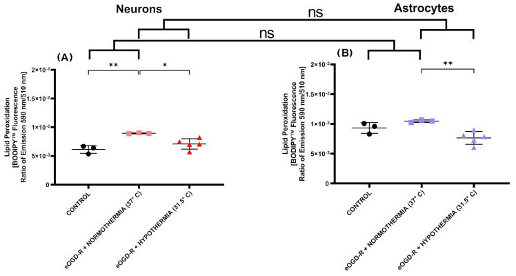
The close interaction between neurons and astrocytes has been extensively studied. However, the specific behavior of these cells after ischemia-reperfusion injury and hypothermia remains poorly characterized. A growing body of evidence suggests that mitochondria function and putative transference between neurons and astrocytes may play a fundamental role in adaptive and homeostatic responses after systemic insults such as cardiac arrest, which highlights the importance of a better understanding of how neurons and astrocytes behave individually in these settings. Brain injury is one of the most important challenges in post-cardiac arrest syndrome, and therapeutic hypothermia remains the single, gold standard treatment for neuroprotection after cardiac arrest. In our study, we modeled ischemia-reperfusion injury by using in vitro enhanced oxygen-glucose deprivation and reperfusion (eOGD-R) and subsequent hypothermia (HPT) (31.5 °C) to cell lines of neurons (HT-22) and astrocytes (C8-D1A) with/without hypothermia. Using cell lysis (LDH; lactate dehydrogenase) as a measure of membrane integrity and cell viability, we found that neurons were more susceptible to eOGD-R when compared with astrocytes. However, they benefited significantly from HPT, while the HPT effect after eOGD-R on astrocytes was negligible. Similarly, eOGD-R caused a more significant reduction in adenosine triphosphate (ATP) in neurons than astrocytes, and the ATP-enhancing effects from HPT were more prominent in neurons than astrocytes. In both neurons and astrocytes, measurement of reactive oxygen species (ROS) revealed higher ROS output following eOGD-R, with a non-significant trend of differential reduction observed in neurons. HPT after eOGD-R effectively downregulated ROS in both cells; however, the effect was significantly more effective in neurons. Lipid peroxidation was higher after eOGD-R in neurons, while in astrocytes, the increase was not statistically significant. Interestingly, HPT had similar effects on the reduction in lipoperoxidation after eOGD-R with both types of cells. While glutathione (GSH) levels were downregulated after eOGD-R in both cells, HPT enhanced GSH in astrocytes, but worsened GSH in neurons. In conclusion, neuron and astrocyte cultures respond differently to eOGD-R and eOGD-R + HTP treatments. Neurons showed higher sensitivity to ischemia-reperfusion insults than astrocytes; however, they benefited more from HPT therapy. These data suggest that given the differential effects from HPT in neurons and astrocytes, future therapeutic developments could potentially enhance HPT outcomes by means of neuronal and astrocytic targeted therapies.

Keywords: astrocytes; cardiac arrest; hypothermia; ischemia-reperfusion injury; neurons; neuroprotection.


Conflict of interest statement

The authors declare that the research was conducted in the absence of any commercial or financial relationships that could be construed as a potential conflict of interest.

Figures

Figure 1
Figure 1
(A) In neurons, 6 h ischemia and 20 h reperfusion protocol in vitro (eOGD-R) caused significant cell lysis as measured by LDH (p-value ≤ 0.0001). Hypothermia (31.5 °C) treatment for 20 h significantly protected neurons against eOGD-R induced cell lysis, as shown by LDH downregulation (p-value ≤ 0.0001). (B) In astrocytes, 6 h + 20 h eOGD-R caused significant cell lysis as measured by LDH (p-value ≤ 0.0001). Hypothermia (31.5 °C) treatment for 20 h did not cause any significant effect on astrocytes against eOGD-R induced cell lysis (p-value = 0.86). Overall, neurons showed higher susceptibility to ischemia-reperfusion insults versus astrocytes (p-value ≤ 0.0001); however, only neurons showed cytoprotection from hypothermia treatment (p-value ≤ 0.0001).
Figure 2
Figure 2
(A) In neurons, 6 h ischemia and 20 h reperfusion protocol in vitro (eOGD-R) caused significant ATP depletion versus the control (p-value ≤ 0.0001). Hypothermia (31.5 °C) treatment significantly protected neurons against eOGD-R insult, as shown by higher ATP levels compared with the normothermia group (p-value ≤ 0.0001). Units expressed as relative light units (RLUs). (B) In astrocytes, 6 h + 20 h normothermia eOGD-R caused significant ATP depletion versus the controls (p-value ≤ 0.0001). Hypothermia (31.5 °C) treatment for 20 h significantly protected astrocytes against eOGD-R insult, as shown by higher ATP levels compared with the normothermia group (p-value ≤ 0.0001). Compared with astrocytes, neurons presented the highest susceptibility for ATP depletion after eOGD-R (p-value ≤ 0.0001); however, hypothermia ameliorated ATP deficit most consistently in neurons (p-value ≤ 0.0001). Units expressed as RLU.
Figure 3
Figure 3
(A) In neurons, normothermic 6 h ischemia and 20 h reperfusion protocol in vitro (eOGD-R) caused significant reactive oxygen species (ROS) production versus the control (p-value ≤ 0.0001). Hypothermia treatment (31.5 °C) for 20 h remarkably prevented ROS burst against eOGD-R insult, as shown by ROS downregulation compared with the normothermia group (p-value ≤ 0.0001). (B) In astrocytes, eOGD-R caused significant ROS production versus the control (p-value ≤ 0.01). Hypothermia treatment (31.5 °C) remarkably prevented ROS burst against eOGD-R insult, as shown by ROS downregulation compared with the normothermia group (p-value ≤ 0.0001). Compared with astrocytes, neurons had a non-significant positive trend on ROS levels after normothermia eOGD-R ((A,B) p-value = 0.17), while hypothermia in neurons after eOGD-R resulted in drastic ROS downregulation. In astrocytes, hypothermia after eOGD-R caused, on average, lower ROS production than its control, but was not statistically significant (p-value = 0.09). Hypothermia had a more evident effect downregulating ROS generation in neurons compared to astrocytes (p ≤ 0.0001).
Figure 4
Figure 4
(A) In neurons, normothermic 6 h ischemia and 20 h reperfusion protocol in vitro (eOGD-R) caused significant lipid peroxidation (LPO) output versus the control (p-value ≤ 0.01). Hypothermia treatment (31.5 °C) for 20 h downregulated LPO generation compared to the normothermia group following eOGD-R insult (p-value ≤ 0.05). (B) In astrocytes, 6 h + 20 h eOGD-R did not cause a statistically significant increase in LPO production (p-value = 0.31). Hypothermia treatment (31.5 °C) for 20 h significantly downregulated LPO after eOGD-R (p-value ≤ 0.01). Of note, hypothermia in astrocytes after eOGD-R decreased LPO at average values below the control, but was not statistically significant (p-value = 0.0790). In conclusion, LPO was significantly upregulated after normothermic eOGD-R in neurons but not in astrocytes. In both cells, hypothermia after eOGD-R significantly downregulated LPO compared with the normothermia eOGD-R groups. Hypothermic eOGD-R downregulated LPO in astrocytes, causing on average a non-significant trend below its control. No statistically significant differential effect was observed on how hypothermia after eOGD-R affected LPO output in neurons and astrocytes (p-value = 0.27).
Figure 5
Figure 5
(A) In neurons, normothermic 6 h ischemia and 20 h reperfusion protocol in vitro (eOGD-R) caused significant depletion of glutathione (GSH) output versus the control (p-value ≤ 0.0001). Hypothermia treatment (31.5 °C) for 20 h further decreased GSH production versus the controls and normothermia eOGD-R (p-value ≤ 0.0001 and ≤ 0.001, respectively). Units expressed as relative light units (RLUs). (B) In astrocytes, 6 h + 20 h normothermia eOGD-R significantly downregulated the GSH levels (p-value ≤ 0.0001). Therapeutic hypothermia (31.5 °C) for 20 h significantly upregulated GSH versus normothermia eOGD-R (p-value ≤ 0.01). Although no differential effect was observed between neurons and astrocytes following eOGD-R, hypothermia treatment induced a significant and contrasting effect on GSH production (p-value ≤ 0.0001). Units expressed as RLU.

Similar articles

Cited by

References

    1. Hayashi M., Shimizu W., Albert C.M. The spectrum of epidemiology underlying sudden cardiac death. Circ. Res. 2015;116:1887–1906. doi: 10.1161/CIRCRESAHA.116.304521. - DOI - PMC - PubMed
    1. Elmer J., Callaway C.W. The Brain after Cardiac Arrest. Semin. Neurol. 2017;37:19–24. doi: 10.1055/s-0036-1597833. - DOI - PMC - PubMed
    1. Sekhon M.S., Ainslie P.N., Griesdale D.E. Clinical pathophysiology of hypoxic ischemic brain injury after cardiac arrest: A ‘two-hit’ model. Crit. Care. 2017;21:90. doi: 10.1186/s13054-017-1670-9. - DOI - PMC - PubMed
    1. Honda H.M., Korge P., Weiss J.N. Mitochondria and Ischemia/Reperfusion Injury. Ann. N. Y. Acad. Sci. 2005;1047:248–258. doi: 10.1196/annals.1341.022. - DOI - PubMed
    1. Nakamura Y., Lo E.H., Hayakawa K. Placental Mitochondria Therapy for Cerebral Ischemia-Reperfusion Injury in Mice. Stroke. 2020;51:3142–3146. doi: 10.1161/STROKEAHA.120.030152. - DOI - PMC - PubMed

LinkOut - more resources