. 2024 Aug 9;12(8):1814.
doi: 10.3390/biomedicines12081814.

Cardiomyocyte Regeneration in Human Myocarditis

Affiliations

Cardiomyocyte Regeneration in Human Myocarditis

Andrea Frustaci et al. Biomedicines. .

Abstract

Background: Newly generated cardiomyocytes (NGCs) concur with the recovery of human myocarditis occurring spontaneously in around 50% of cases. However, NGCs decline with age, and their modality of myocardial homing and integration are still unclear.

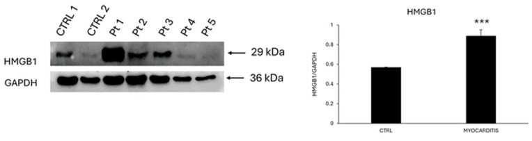
Methods: We retrospectively assessed NGCs in 213 consecutive patients with endomyocardial biopsy denoting acute myocarditis, with normal coronaries and valves. Tissue samples were processed for histology (H&E), immunohistochemistry for the evaluation of inflammatory infiltrates, immunostaining for alpha-sarcomeric-actin, junctional connexin-43, Ki-67, and phosphorylated STAT3 (p-STAT3), and Western blot (WB) for HMGB1. Frozen samples were analyzed using polymerase chain reaction (PCR) for cardiotropic viruses. Controls included 20 normal surgical biopsies.

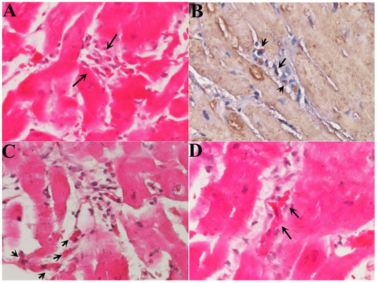
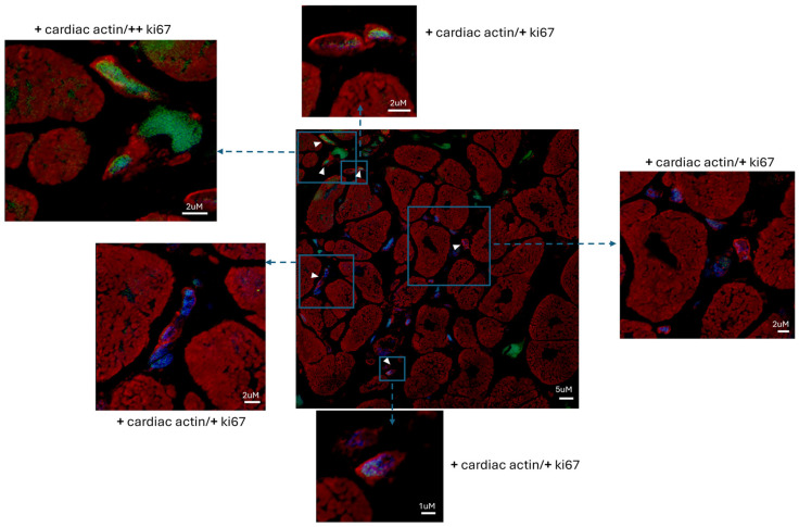
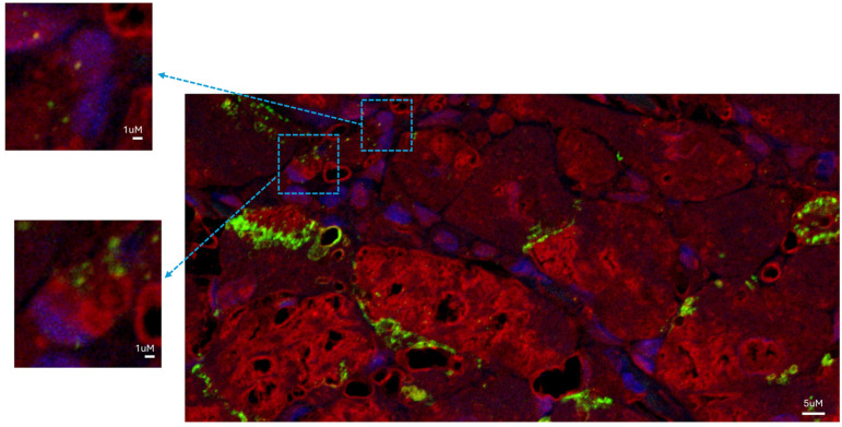
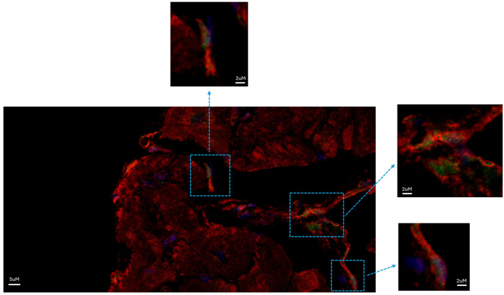
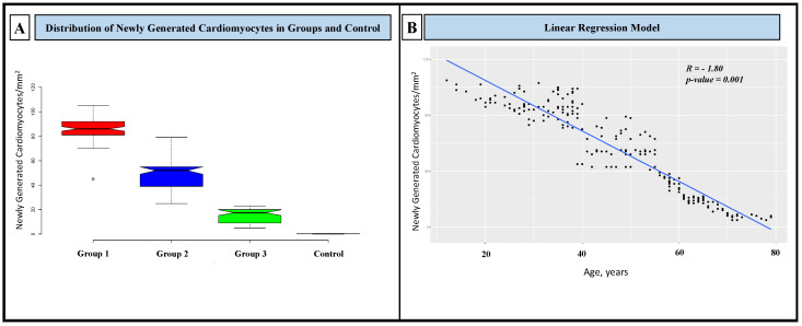
Results: NGCs were defined as small myocytes (diameter < 10 µm) with nuclear positivity to Ki-67 and p-STAT3 and positive immunostaining for cytoplasmic α-sarcomeric actin and connexin-43. Their number/mm2 in relation to age and pathway of integration was evaluated. NGCs crossed the membrane and grew integrated within the empty necrotic myocytes. NGC mean diameter was 6.6 ± 3.34 vs. 22.5 ± 3.11 µm adult cells; their number, in comparison to LVEF, was 86.3 ± 10.3/mm2 in patients between 18 and 40 years, 50.4 ± 13.8/mm2 in those between 41 and 60, and 15.1 ± 5.7/mm2 in those between 61 and 80. Control NGCs' mean diameter was 0.2 ± 0.2 mm2. PCR was positive for viral genomes in 16% of cases; NGCs were not statistically different in viral and non-viral myocarditis. WB analysis revealed a higher expression of HMGB1 in myocarditis compared to myocardial controls.

Conclusions: NGCs are constantly recognizable in acute human myocarditis. Their number declines with age. Their integration within necrotic myocytes allows for the preservation of the cardiac structure and function.

Keywords: STAT3; myocarditis; newly generated cardiomyocytes.


Conflict of interest statement

The authors declare that they have no competing interests.

Figures

Figure 1
Figure 1
Homing and integration of NGCs into inflamed myocardium. (A) Several cells resembling small myocytes (arrows) approach an area of cardiomyocyte necrosis caused by lymphocytic myocarditis (H&E 400×). (B) Immunohistochemistry for alpha-sarcomeric-actin (400×) shows the presence of contractile material in the small cells. (C) Multiple NGCs are lining up (arrows) to enter an empty cardiomyocyte, crossing its cell membrane (H&E 400×). (D) NGCs (arrows) are growing inside of a cardiomyocyte that has undergone a myocytolytic process (H&E, 400×).
Figure 2
Figure 2
Myocarditis induces the formation of proliferating NGCs’ α-sarcomeric-expressing cells in the human left ventricle. Newly formed cells positive for α-sarcomeric actin (red fluorescence) expressed Ki-67 (green fluorescence), as indicated by the arrowheads. Red fluorescence indicates α-sarcomeric actin immunostaining; green fluorescence indicates Ki-67; blue fluorescence indicates DAPI staining of nuclei. Newly formed cells positive for α-sarcomeric actin (red fluorescence) expressed Ki-67 (green fluorescence), as indicated by the arrowheads. Bar = 5 µm (inserts: magnified version; bar = 2 µm or 1 µm).
Figure 3
Figure 3
Newly generated cardiomyocytes in the human left ventricle were functionally competent. NGCs expressed connexin-43, detected as punctuate staining (green fluorescence) in the gap-junctional regions between cardiomyocytes in the left ventricular tissue. Newly formed α-sarcomeric-expressing cells expressed connexin-43. Red fluorescence indicates α-sarcomeric actin immunostaining; green fluorescence indicates connexin-43; blue fluorescence indicates DAPI staining of nuclei. Bar = 5 µm (inserts: magnified version; bar = 1 µm).
Figure 4
Figure 4
STAT3 was activated in NGCs of patients diagnosed with myocarditis. NGCs expressed pSTAT3 in the nuclei (green fluorescence). Red fluorescence indicates α-sarcomeric actin immunostaining; green fluorescence indicates pSTAT3; blue fluorescence indicates DAPI staining of nuclei. Bar = 5 μm (inserts: magnified version; bar = 2 µm).
Figure 5
Figure 5
HMGB1 expression is up-regulated in myocarditis. Western blot analysis showing the expression of HMGB1 in LV endomyocardial biopsies of myocarditis compared to controls (CTRL). The same filter was probed with anti-GAPDH pAb to show equal loading. Left panel: a representative Western blotting of three replicates is shown. Marker: lane 1 = CTRL 1, lane 2 = CTRL 2, lane 3 = pt 1, lane 4 = pt 2, lane 5 = pt 3, lane 6 = pt 4, and lane 7 = pt 5. Right panel: densitometric analysis of Western blot (mean values) from CTRL patients (n = 2) and patients with myocarditis (n = 5). Data are shown as means ± SEM. ***, p < 0.001.
Figure 6
Figure 6
Distribution of newly generated cardiomyocytes in groups and controls and their correlation with age. (A) NGCs were statistically different in the three groups and between groups and controls (p-value between groups: 0.001). In the box plot are represented the median (line in the middle) and the middle “box” (1st–3rd quartile) of all values. The upper and lower whiskers represent scores outside of the middle 50%. (B) NGCs linearly decreased with age.

Similar articles

References

    1. D’Ambrosio A. The Fate of Acute Myocarditis between Spontaneous Improvement and Evolution to Dilated Cardiomyopathy: A Review. Heart. 2001;85:499–504. doi: 10.1136/heart.85.5.499. - DOI - PMC - PubMed
    1. Frustaci A., Chimenti C., Pieroni M., Salvatori L., Morgante E., Sale P., Ferretti E., Petrangeli E., Gulino A., Russo M.A. Cell Death, Proliferation and Repair in Human Myocarditis Responding to Immunosuppressive Therapy. Mod. Pathol. 2006;19:755–765. doi: 10.1038/modpathol.3800594. - DOI - PubMed
    1. Miyawaki A., Obana M., Mitsuhara Y., Orimoto A., Nakayasu Y., Yamashita T., Fukada S., Maeda M., Nakayama H., Fujio Y. Adult Murine Cardiomyocytes Exhibit Regenerative Activity with Cell Cycle Reentry through STAT3 in the Healing Process of Myocarditis. Sci. Rep. 2017;7:1407. doi: 10.1038/s41598-017-01426-8. - DOI - PMC - PubMed
    1. Han C., Nie Y., Lian H., Liu R., He F., Huang H., Hu S. Acute Inflammation Stimulates a Regenerative Response in the Neonatal Mouse Heart. Cell Res. 2015;25:1137–1151. doi: 10.1038/cr.2015.110. - DOI - PMC - PubMed
    1. Fang Y., Gupta V., Karra R., Holdway J.E., Kikuchi K., Poss K.D. Translational Profiling of Cardiomyocytes Identifies an Early Jak1/Stat3 Injury Response Required for Zebrafish Heart Regeneration. Proc. Natl. Acad. Sci. USA. 2013;110:13416–13421. doi: 10.1073/pnas.1309810110. - DOI - PMC - PubMed

LinkOut - more resources