. 2024 Aug 12;12(8):1835.
doi: 10.3390/biomedicines12081835.

Surface Pre-Reacted Glass-Ionomer Eluate Suppresses Osteoclastogenesis through Downregulation of the MAPK Signaling Pathway

Affiliations

Surface Pre-Reacted Glass-Ionomer Eluate Suppresses Osteoclastogenesis through Downregulation of the MAPK Signaling Pathway

Janaki Chandra et al. Biomedicines. .

Abstract

Surface pre-reacted glass-ionomer (S-PRG) is a new bioactive filler utilized for the restoration of decayed teeth by its ability to release six bioactive ions that prevent the adhesion of dental plaque to the tooth surface. Since ionic liquids are reported to facilitate transepithelial penetration, we reasoned that S-PRG applied to root caries could impact the osteoclasts (OCs) in the proximal alveolar bone. Therefore, this study aimed to investigate the effect of S-PRG eluate solution on RANKL-induced OC-genesis and mineral dissolution in vitro. Using RAW264.7 cells as OC precursor cells (OPCs), TRAP staining and pit formation assays were conducted to monitor OC-genesis and mineral dissolution, respectively, while OC-genesis-associated gene expression was measured using quantitative real-time PCR (qPCR). Expression of NFATc1, a master regulator of OC differentiation, and the phosphorylation of MAPK signaling molecules were measured using Western blotting. S-PRG eluate dilutions at 1/200 and 1/400 showed no cytotoxicity to RAW264.7 cells but did significantly suppress both OC-genesis and mineral dissolution. The same concentrations of S-PRG eluate downregulated the RANKL-mediated induction of OCSTAMP and CATK mRNAs, as well as the expression of NFATc1 protein and the phosphorylation of ERK, JNK, and p38. These results demonstrate that S-PRG eluate can downregulate RANKL-induced OC-genesis and mineral dissolution, suggesting that its application to root caries might prevent alveolar bone resorption.

Keywords: S-PRG; TRAP staining; bioactive filler; hydroxyapatite; osteoclast.


Conflict of interest statement

All authors, except T.N., declare no conflicts of interest. T.N. is a member of Shifu inc. which provided the S-PRG materials. Shofu Inc. has been sharing S-PRG filler and S-PGR eluate to all researchers worldwide free of charge. T.N.’s role in this study was limited to providing high-quality S-PGR eluate and developing the research hypothesis together with corresponding authors in strict compliance with conflict of interest guidelines.

Figures

Figure 1
Figure 1
Timeline for release assay. S-PRG eluate was applied to HA-coated plate for 24 h. Fresh media were added to the wells, and the supernatant was collected at different time points. Collected supernatant was applied to OC-genesis assay.
Figure 2
Figure 2
Cytotoxicity of S-PRG eluate. S-PRG eluate had no cytotoxic effect on RAW264.7 cells after diluting to more than 1/200. The horizontal dashed line with a value of 100 indicates the survival rate of RAW264.7 cells cultured in the absence of S-PRG eluate. Results were presented as the means ± SD. *: p < 0.05, vs. control without S-PRG eluate.
Figure 3
Figure 3
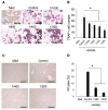
Evaluation of OC-genesis. The number of TRAP-positive cells by adding S-PRG eluate to osteoclast precursors (A,B). Quantification of pit area when adding S-PRG eluate. S-PRG eluate significantly decreased pit area created by osteoclasts compared to control (C,D). Scale bars indicate 100 µm. Results were presented as the means ± SD. *: p < 0.05, vs. control.
Figure 4
Figure 4
Quantification of mRNA expression related to OC-genesis (A) and NFATc1 induction (B,C) when adding S-PRG eluate. Results were presented as the means ± SD. *: p < 0.05, vs. control.
Figure 5
Figure 5
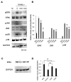
Phosphorylation of MAPK proteins (A,B) and the expression of total IκBα (C,D) by adding RANKL and S-PRG eluate. Results were presented as the means ± SD. *: p < 0.05, vs. control.
Figure 6
Figure 6
MAPK inhibitors suppressed the OC-genesis. The inhibitors against ERK, p38, and JNK significantly decreased the number of TRAP-positive cells (A,B) and pit area (C,D). Scale bars indicate 100 µm. Results were presented as the means ± SD. *: p < 0.05, vs. control.
Figure 7
Figure 7
S-PRG eluate, in combination with MAPK inhibitors, suppressed the OC-genesis. The addition of S-PRG elute to inhibitors against ERK, p38, and JNK adding to S-PRG eluate significantly decreased the number of TRAP-positive cells. Results were presented as the means ± SD. Scale bars indicate 100 µm. *: p < 0.05, vs. control.
Figure 8
Figure 8
Evaluation of the number of TRAP-positive cells (A,B) when adding S-PRG eluate released from HA. Scale bars indicate 100 µm. Results were presented as the means ± SD. *: p < 0.05, vs. control.
Figure 9
Figure 9
Each ion present in S-PRG eluate inhibited RANKL-induced OC-genesis. Sr2+, Al3+, F, and B3+ significantly decreased the number of TRAP-positive cells induced in Raw264.7 cells stimulated with RANKL (A,B). Pit area created by Raw264.7 cells incubated with Sr2+, F, and B3+ in the presence of RANKL was smaller than that in control (C,D). Scale bars indicate 100 µm. Results were presented as the means ± SD. *: p < 0.05, vs. control.
Figure 10
Figure 10
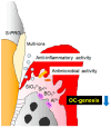
Potential effect of S-PRG eluate on periodontal disease. S-PRG eluate downregulates OC-genesis, as well as anti-inflammatory activity and antimicrobial activity.

References

    1. Eke P.I., Dye B.A., Wei L. Thornton-Evans GO, Genco RJ. Prevalence of periodontitis in adults in the United States: 2009 and 2010. J. Dent. Res. 2012;91:914–920. doi: 10.1177/0022034512457373. - DOI - PubMed
    1. Kassebaum N.J., Bernabé E., Dahiya M., Bhandari B., Murray C.J., Marcenes W. Global burden of severe periodontitis in 1990-2010: A systematic review and meta-regression. J. Dent. Res. 2014;93:1045–1053. doi: 10.1177/0022034514552491. - DOI - PMC - PubMed
    1. Pihlstrom B.L., Michalowicz B.S., Johnson N.W. Periodontal diseases. Lancet. 2005;366:1809–1820. doi: 10.1016/S0140-6736(05)67728-8. - DOI - PubMed
    1. Bhuyan R., Bhuyan S.K., Mohanty J.N., Das S., Juliana N., Juliana I.F. Periodontitis and Its Inflammatory Changes Linked to Various Systemic Diseases: A Review of Its Underlying Mechanisms. Biomedicines. 2022;10:2659. doi: 10.3390/biomedicines10102659. - DOI - PMC - PubMed
    1. Lalla E., Papapanou P.N. Diabetes mellitus and periodontitis: A tale of two common interrelated diseases. Nat. Rev. Endocrinol. 2011;7:738–748. doi: 10.1038/nrendo.2011.106. - DOI - PubMed

LinkOut - more resources