. 2024 Aug 15;12(8):1856.
doi: 10.3390/biomedicines12081856.

Sensory Neurons Release Cardioprotective Factors in an In Vitro Ischemia Model

Affiliations

Sensory Neurons Release Cardioprotective Factors in an In Vitro Ischemia Model

Clara Hoebart et al. Biomedicines. .

Abstract

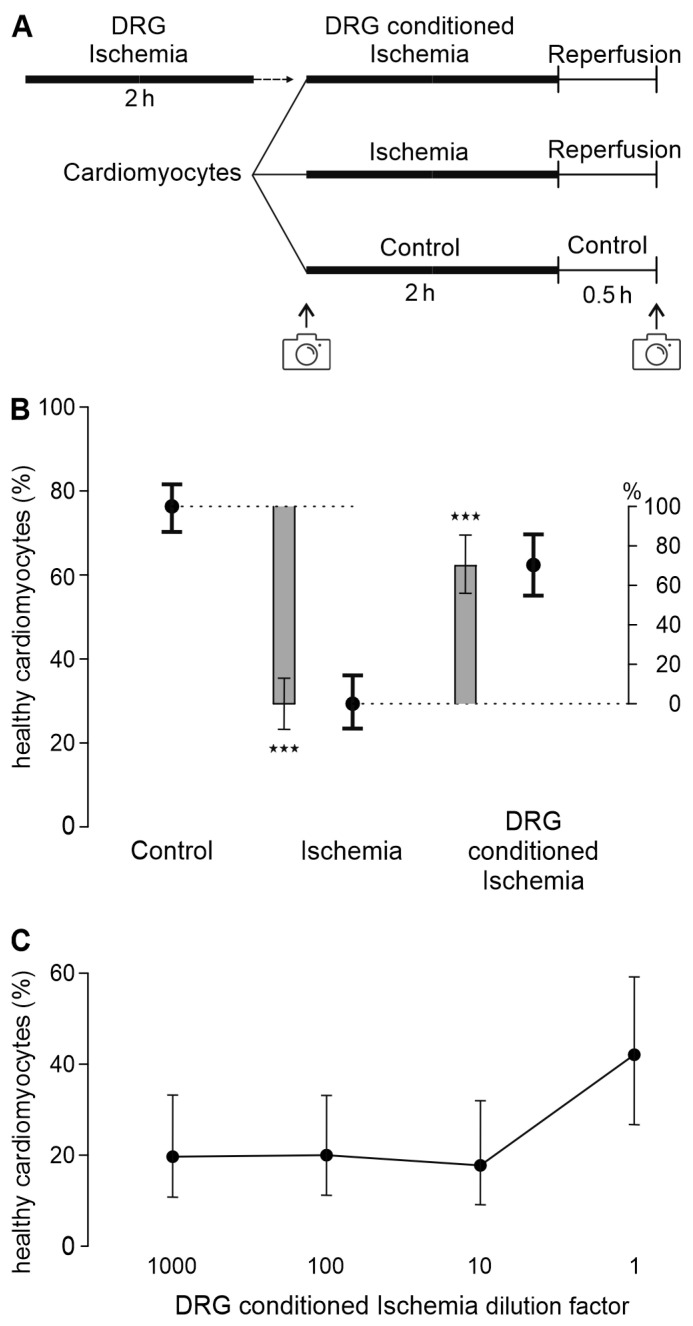
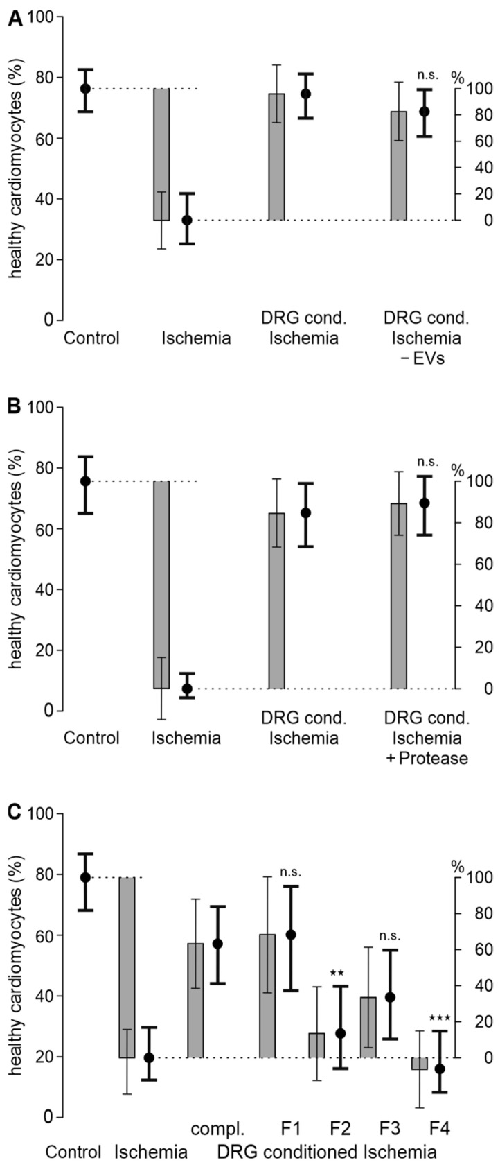
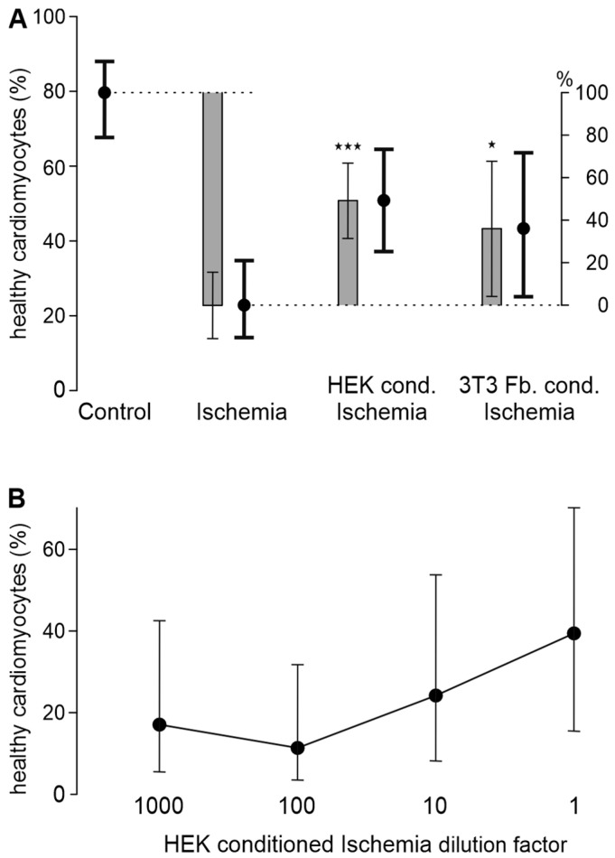
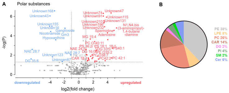
Sensory neurons densely innervate the myocardium. The role of their sensing and response to acute and prolonged ischemia is largely unclear. In a cellular model of ischemia-reperfusion injury, the presence of sensory neurons increases cardiomyocyte survival. Here, after the exclusion of classical neurotransmitter release, and measurement of cytokine release, we modified the experiment from a direct co-culture of primary murine cardiomyocytes and sensory neurons to a transfer of the supernatant. Sensory neurons were exposed to ischemia and the resulting conditioned supernatant was transferred onto cardiomyocytes. This approach largely increased the tolerance of cardiomyocytes to ischemia and reperfusion. Towards the identification of the mechanism, it was demonstrated that after ten-fold dilution, the conditioned solution lost its protective effect. The effect remained after removal of extracellular vesicles by ultracentrifugation, and was not affected by exposure to protease activity, and fractionation pointed towards a hydrophilic agent. Solutions conditioned by HEK293t cells or 3T3 fibroblasts also increase cardiomyocyte survival, but to a lower degree. A metabolomic search identified 64 at least two-fold changed metabolites and lipids. Many of these could be identified and are involved in essential cellular functions. In the presented model for ischemia-reperfusion, sensory neurons secrete one or more cardioprotective substances that can improve cardiomyocyte survival.

Keywords: cardiomyocytes; ischemia; ischemia-reperfusion; sensory neurons.


Conflict of interest statement

The authors declare no conflicts of interest. The funders had no role in the design of the study; in the collection, analyses, or interpretation of data; in the writing of the manuscript; or in the decision to publish the results.

Figures

Figure 1
Figure 1
DRG exposed to ischemia release transferable mediators protecting cardiomyocytes from ischemia-reperfusion injury. (A) Experimental protocol of transfer of DRG-conditioned ischemic solution to cardiomyocytes in in vitro ischemia-reperfusion model. (B) Estimated survival probabilities for control, ischemia-reperfusion conditions, and treatment with DRG-conditioned ischemic solution are shown as black dots ± 95% CI. Estimated differences are shown as gray bars ± 95% CI. Cardiomyocytes and DRG were each derived from n = 24 animals, respectively. The dotted lines serves as visual reference. Cardiomyocytes of each animal were distributed to three experimental conditions, and DRG of animal sacrificed before were used to prepare conditioned ischemic solution. Data of all respective experiments are plotted in Figure S3A, ★★★ p < 0.001. (C) For all dilutions of DRG-conditioned ischemic solutions, increase in cardiomyocyte survival is less than that for undiluted solution (p < 0.001 each). Estimated survival probabilities for each concentration are shown as black dots ± 95% CI, connected by line. Cardiomyocytes were derived from n = 3 animals; DRG for DRG-conditioned ischemia solution were also derived from n = 3 animals. Cardiomyocytes of each animal were distributed to four experimental conditions. Data separated by experimental day or per well are presented in Supplementary Figure S3.
Figure 2
Figure 2
The improvement in cardiomyocyte survival by the DRG-conditioned ischemic solution is not mediated by extracellular vesicles or proteins, but rather by one or more hydrophilic mediator(s). (A) Cardiomyocytes were treated with the DRG-conditioned ischemic solution with and without extracellular vesicles (EVs, removed by ultracentrifugation). p = 0.21 vs. DRG-conditioned ischemia. Cardiomyocytes were derived from n = 3 animals; DRG for the DRG-conditioned ischemia solution were also derived from n = 3 animals. Cardiomyocytes of each animal were distributed to all 4 experimental conditions. (B) Cardiomyocytes were treated with the DRG-conditioned ischemic solution, and pretreated with protease or not pretreated. p = 0.65 vs. DRG-conditioned ischemia. Cardiomyocytes were derived from n = 3 animals; DRG for the DRG-conditioned ischemia solution were also derived from n = 3 animals. Cardiomyocytes of each animal were distributed to all 4 experimental conditions. (C) Cardiomyocytes were treated with the complete DRG-conditioned ischemic solution (compl.) or with 4 different fractions, F1–F4, of the DRG-conditioned ischemic solution, which were obtained using a C18 column and different eluents. Fraction 1 was eluted by water, and fractions 2–4 by 30%, 60%, and 100% acetonitrile, respectively. In all panels, estimated survival probabilities for each group are shown as black dots ± 95% CI and estimated differences are shown as gray bars ± 95% CI. The dotted lines serves as visual reference. p-Values refer to comparisons of respective fractions with DRG-conditioned ischemia, ★★ p = 0.01 and ★★★ p < 0.001. n.s.: not significant. Cardiomyocytes were derived from n = 3 animals; DRG for the DRG-conditioned ischemia solution were also derived from n = 3 animals. Data for each well are presented in Figure S4E.
Figure 3
Figure 3
Certain cell lines can increase cardiomyocyte survival in ischemia-reperfusion. (A) The cell lines HEK293t and 3T3 fibroblasts (Fb.) conditioned the ischemic solution before it was applied to cardiomyocytes. Estimated survival probabilities for each cell line are shown as black dots ± 95% CI and estimated differences are shown as gray bars ± 95% CI. The dotted lines serves as visual reference. ★ p = 0.027 and ★★★ p < 0.001 vs. ischemia. Cardiomyocytes were derived from 4 to 10 animals. The exact number of animals per condition is visualized in Figure S5A. (B) The effect of the HEK293t-conditioned ischemic solution is concentration-dependent, with dilutions by a factor of 1000, 100, and 10 shown. Estimated survival probabilities for each concentration are shown as black dots ± 95% CI, connected by a line. Data are derived from n = 3 animals killed on separate experimental days. Data from each experimental day are shown in Figure S5B; data from each well are depicted in Figure S5C.
Figure 4
Figure 4
Regulated metabolites and lipids in ischemia-conditioned DRG. (A) A volcano plot with equal group variance showing the relative regulation in the DRG-conditioned ischemic solution compared to the reference DRG-conditioned external solution (p-value cut-off at 0.05, –logP(0.05) = 1.30, fold change cut-off = 2). Substances that could not be identified using reference libraries or putative annotation were labeled as ‘unknown’ and enumerated. (B) A pie chart of percentages of different categories of lipids found. Phosphatidylethanolamines (PEs), lysophosphatidylethanolamines (LPEs), phosphatidylcholines (PCs), acylcarnitines (CARs), diacylglycerols (DGs), phosphatidylinositol (PI), sphingomyelin (SM), and ceramides (Cer). The analysis was based on the pooled solutions generated by n = 9 animals per condition.

Similar articles

References

    1. Roth G.A., Mensah G.A., Johnson C.O., Addolorato G., Ammirati E., Baddour L.M., Barengo N.C., Beaton A.Z., Benjamin E.J., Benziger C.P., et al. Global Burden of Cardiovascular Diseases and Risk Factors, 1990–2019: Update from the GBD 2019 Study. J. Am. Coll. Cardiol. 2020;76:2982–3021. doi: 10.1016/j.jacc.2020.11.010. - DOI - PMC - PubMed
    1. Thygesen K., Alpert J.S., Jaffe A.S., Chaitman B.R., Bax J.J., Morrow D.A., White H.D. Fourth Universal Definition of Myocardial Infarction (2018) J. Am. Coll. Cardiol. 2018;72:2231–2264. doi: 10.1016/j.jacc.2018.08.1038. - DOI - PubMed
    1. Ibanez B., James S., Agewall S., Antunes M.J., Bucciarelli-Ducci C., Bueno H., Caforio A.L.P., Crea F., Goudevenos J.A., Halvorsen S., et al. 2017 ESC Guidelines for the management of acute myocardial infarction in patients presenting with ST-segment elevation: The Task Force for the management of acute myocardial infarction in patients presenting with ST-segment elevation of the European Society of Cardiology (ESC) Eur. Heart J. 2018;39:119–177. doi: 10.1093/eurheartj/ehx393. - DOI - PubMed
    1. Cowled P., Fitridge R. In: Mechanisms of Vascular Disease: A Reference Book for Vascular Specialists. Fitridge R., Thompson M., editors. University of Adelaide Press; Adelaide, Australia: 2011. - PubMed
    1. Sanada S., Komuro I., Kitakaze M. Pathophysiology of myocardial reperfusion injury: Preconditioning, postconditioning, and translational aspects of protective measures. Am. J. Physiol. Heart Circ. Physiol. 2011;301:H1723–H1741. doi: 10.1152/ajpheart.00553.2011. - DOI - PubMed

LinkOut - more resources