. 2024 Aug 22;12(8):1927.
doi: 10.3390/biomedicines12081927.

Combination of Anti-CD40 and Anti-CD40L Antibodies as Co-Stimulation Blockade in Preclinical Cardiac Xenotransplantation

Affiliations

Combination of Anti-CD40 and Anti-CD40L Antibodies as Co-Stimulation Blockade in Preclinical Cardiac Xenotransplantation

Martin Bender et al. Biomedicines. .

Abstract

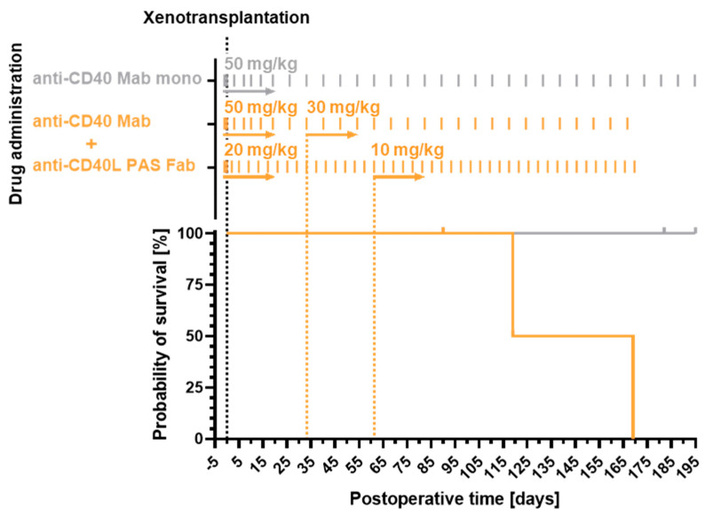
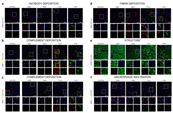
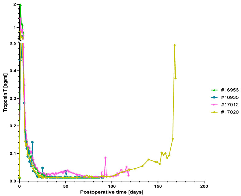
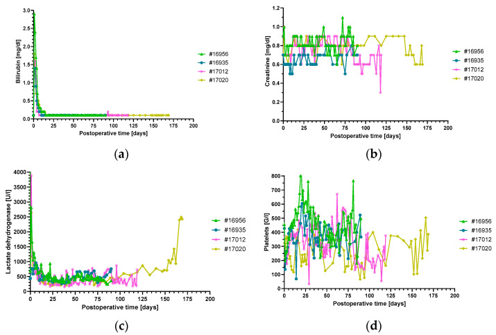
The blockade of the CD40/CD40L immune checkpoint is considered essential for cardiac xenotransplantation. However, it is still unclear which single antibody directed against CD40 or CD40L (CD154), or which combination of antibodies, is better at preventing organ rejection. For example, the high doses of antibody administered in previous experiments might not be feasible for the treatment of humans, while thrombotic side effects were described for first-generation anti-CD40L antibodies. To address these issues, we conducted six orthotopic pig-to-baboon cardiac xenotransplantation experiments, combining a chimeric anti-CD40 antibody with an investigational long-acting PASylated anti-CD40L Fab fragment. The combination therapy effectively resulted in animal survival with a rate comparable to a previous study that utilized anti-CD40 monotherapy. Importantly, no incidence of thromboembolic events associated with the administration of the anti-CD40L PAS-Fab was observed. Two experiments failed early because of technical reasons, two were terminated deliberately after 90 days with the baboons in excellent condition and two were extended to 120 and 170 days, respectively. Unexpectedly, and despite the absence of any clinical signs, histopathology revealed fungal infections in all four recipients. This study provides, for the first time, insights into a combination therapy with anti-CD40/anti-CD40L antibodies to block this immune checkpoint.

Keywords: CD40/CD40L; co-stimulation blockade; heart; orthotopic heart transplantation; xenotransplantation.


Conflict of interest statement

Jan-Michael Abicht, Bruno Reichart, Eckhard Wolf, Paolo Brenner, Arne Skerra and Matthias Längin are founders of XTransplant GmbH. David Ayares is chief executive officer and chief scientific officer of Revivicor, Inc. Michaela Gebauer is an employee, Uli Binder and Arne Skerra are shareholders of XL-protein GmbH. The funders had no role in the design of the study; in the collection, analyses, or interpretation of data; in the writing of the manuscript; or in the decision to publish the results. The other authors declare no conflicts of interest.

Figures

Figure 1
Figure 1
Dosing scheme (top) and survival (bottom) of the study group comprising six animals, which received anti-CD40 IgG4/anti-CD40L PAS-Fab combination therapy (orange). The dosage of the anti-CD40 Mab was reduced within 30 days from initially 50 to 30 mg/kg body weight (bw), and the dosage of the anti-CD40L PAS-Fab was lowered within 60 days from 20 to 10 mg/kg bw. In comparison with a previously reported [4] immunosuppressive regimen with the anti-CD40 Mab alone (gray), there was no significant difference in survival (Log-rank (Mantel–Cox) test, n = 10, p = 0.0896). In the current study group, two technical failures were excluded from the survival analysis. In the previously described group [4], two animals which were positive for PCMV/PRV were excluded. PCMV/PRV, porcine cytomegalovirus/porcine roseolovirus.
Figure 8
Figure 8
Immunofluorescence staining of post-mortem myocardial specimens. Antibody deposition (IgM) (a), complement deposition (C3b/c, C4c, C5b) (b,c), fibrin deposition (Fgn) (d), cardiomyocyte structure (WGA, a lectin staining N-acetyl-D-glucosamine and sialic acid on the cell membrane) (e) and macrophage infiltration (CD68) (f) were analyzed in all donor organs. Scale bar, 100 µm. n = 4 GGTA1-KO, hCD46/hTBM transgenic pigs; n = 1 wild-type pig (control).
Figure 2
Figure 2
Serum troponin T levels. Levels were high after surgery but subsequently dropped to a normal range in animals #16956, #16935 and #17012. The strong increase in serum troponin T levels in animal #17020 indicates myocardial damage due to graft rejection towards the end of the experiment.
Figure 3
Figure 3
Plasma bilirubin (a), creatinine (b) and LDH (c) levels and platelet counts (d), non-suggestive of thrombotic microangiopathy. There were no signs of liver or kidney damage. The increase in LDH in animal #17020 at the end of the experiment was caused by humoral rejection. LDH, lactate dehydrogenase.
Figure 4
Figure 4
Levels of non-Gal-α(1,3)-Gal xenoreactive IgM (a) and IgG (b) as measured by flow cytometry on porcine aortic endothelial cells (PAEC) from GGTA1-KO, hCD46/hTBM transgenic animals. The values from a previously investigated baboon that had rejected an intrathoracic heterotopically transplanted pig heart served as positive control (black) and showed a strong increase in both IgM and IgG, indicative of humoral rejection. Note: while animal #17020 revealed clinical and histological signs of humoral rejection, there was no increase in xenoreactive IgM and IgG.
Figure 5
Figure 5
Cumulative pleural effusions in baboon #17012 (a) and serum levels of the inflammatory markers leukocytes and IL-6 (b), CRP (c) as well as the myocardial markers CK and NT-proBNP (d). The sharp rise in pleural effusions around postoperative day 85 was accompanied by a marked increase in all inflammatory and myocardial markers ((ad) and Figure 2). For comparison, at the beginning of the pleural effusions around postoperative day 30 there was only an increase in IL-6, leukocyte count (b) and also in troponin T levels (Figure 2).
Figure 6
Figure 6
Serum levels of different pro-inflammatory cytokines in all animals of the study group (ag). The marked increase around postoperative day 85 was only observed in baboon #17012 (magenta), whereas #17020 showed elevated levels of some cytokines towards the end of the experiment, when organ rejection occurred. The sharp increase in pleural effusions around postoperative day 85 in baboon #17012 (h) was accompanied by a strong rise in several pro-inflammatory cytokine levels (magenta).
Figure 7
Figure 7
Microscopic findings in post-mortem myocardial specimens. Histological analysis revealed mild-to-marked interstitial edema (arrows) in myocardial specimens of baboons #16956 (a), #16935 (c), #17012 (d) and #17020 (f,h). Fungal thrombi in pulmonal vessels (arrowhead) were detected in all animals (#16956, (b); #17012, (e); #17020, (i); not shown for #16935). Based on capillaritis (arrowhead, (f)), endothelial swelling (arrowhead, (h)) and elongated C4d staining in capillaries (arrows, (g)), baboon #17020 was diagnosed with severe antibody-mediated rejection (AMR3). (ad,f,h,i), H&E staining; (e), Grocott methenamine staining; (g), C4d staining; scale bars = 100 µm.

References

    1. Reichart B., Cooper D.K.C., Langin M., Tonjes R.R., Pierson R.N., Wolf E. Cardiac xenotransplantation: From concept to clinic. Cardiovasc. Res. 2023;118:3499–3516. doi: 10.1093/cvr/cvac180. - DOI - PMC - PubMed
    1. Mohiuddin M.M., Reichart B., Byrne G.W., McGregor C.G. Current status of pig heart xenotransplantation. Pt BInt. J. Surg. 2015;23:234–239. doi: 10.1016/j.ijsu.2015.08.038. - DOI - PMC - PubMed
    1. Längin M., Mayr T., Reichart B., Michel S., Buchholz S., Guethoff S., Dashkevich A., Baehr A., Egerer S., Bauer A., et al. Consistent success in life-supporting porcine cardiac xenotransplantation. Nature. 2018;564:430–433. doi: 10.1038/s41586-018-0765-z. - DOI - PubMed
    1. Reichart B., Längin M., Radan J., Mokelke M., Buttgereit I., Ying J., Fresch A.K., Mayr T., Issl L., Buchholz S., et al. Pig-to-non-human primate heart transplantation: The final step toward clinical xenotransplantation? J. Heart Lung Transplant. 2020;39:751–757. doi: 10.1016/j.healun.2020.05.004. - DOI - PubMed
    1. Mohiuddin M.M., Goerlich C.E., Singh A.K., Zhang T., Tatarov I., Lewis B., Sentz F., Hershfeld A., Braileanu G., Odonkor P., et al. Progressive genetic modifications of porcine cardiac xenografts extend survival to 9 months. Xenotransplantation. 2022;29:e12744. doi: 10.1111/xen.12744. - DOI - PMC - PubMed