. 2024 Aug 6;13(16):4602.
doi: 10.3390/jcm13164602.

Surgical Navigation and CAD-CAM-Designed PEEK Prosthesis for the Surgical Treatment of Facial Intraosseous Vascular Anomalies

Affiliations

Surgical Navigation and CAD-CAM-Designed PEEK Prosthesis for the Surgical Treatment of Facial Intraosseous Vascular Anomalies

Alicia Dean et al. J Clin Med. .

Abstract

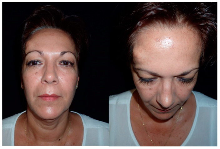
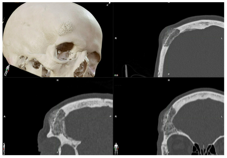
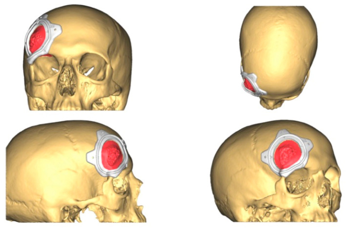
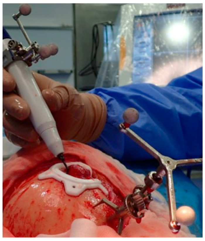
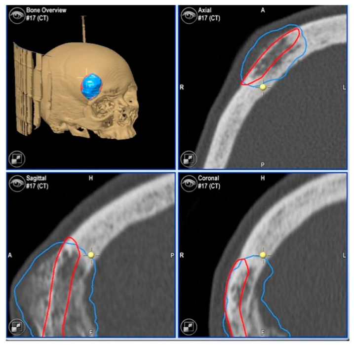
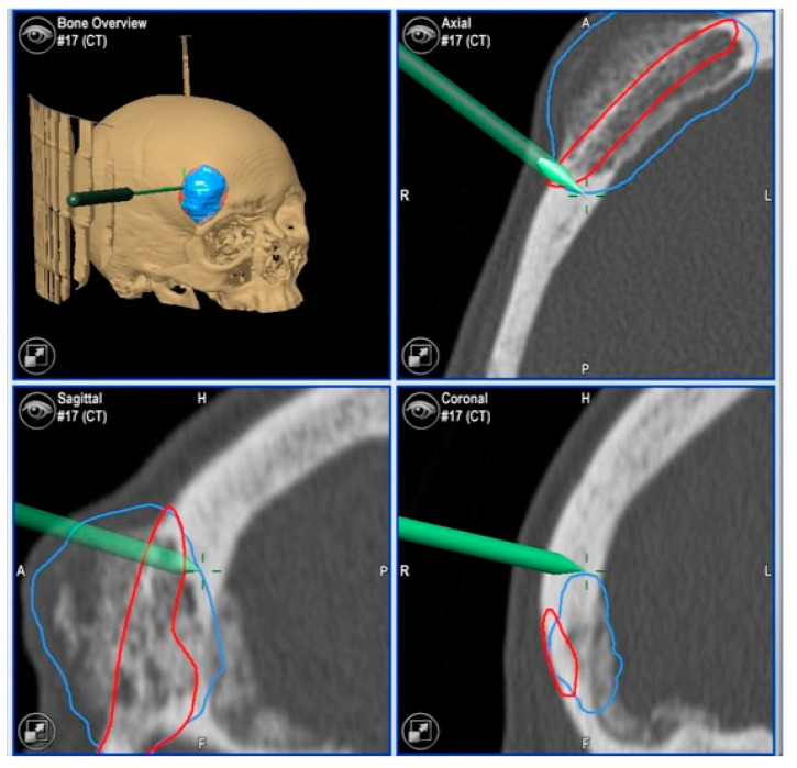
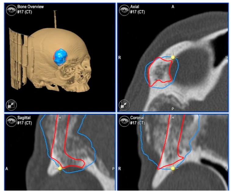
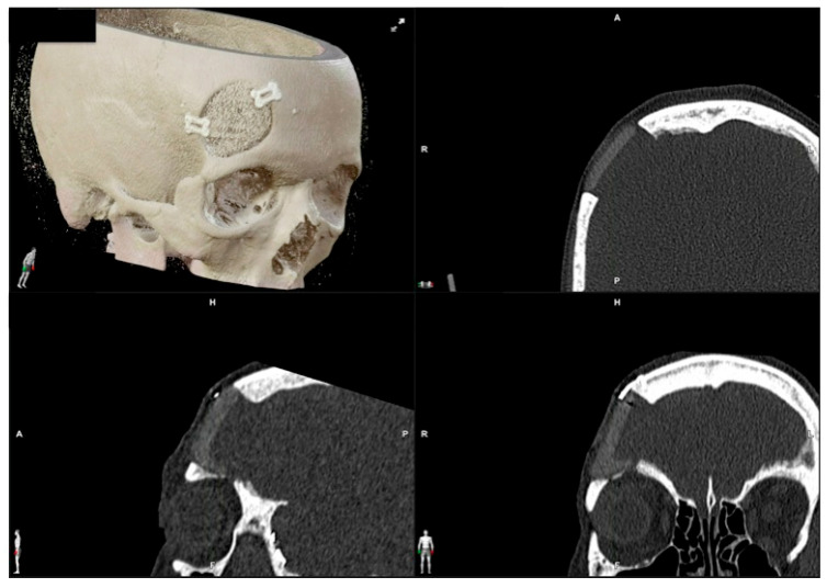
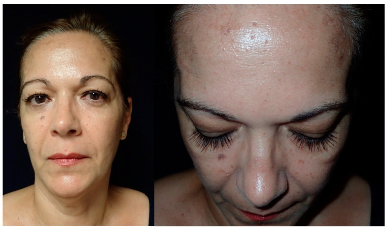
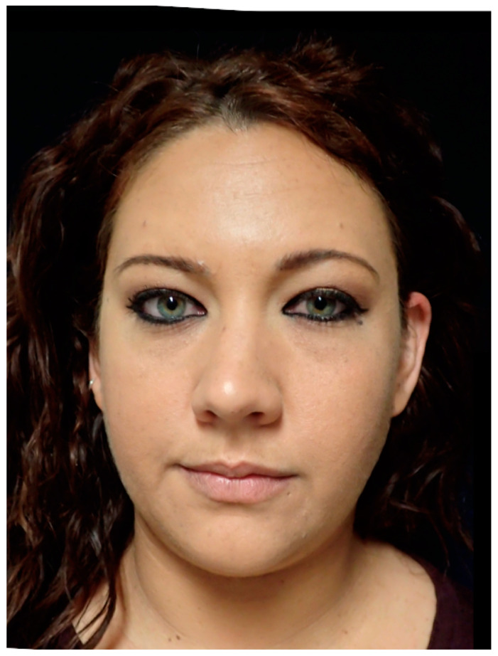
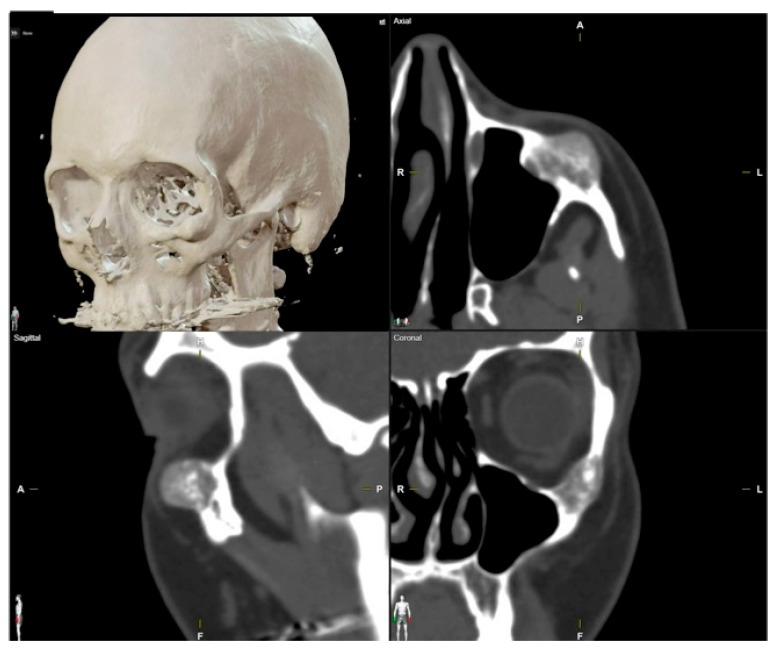
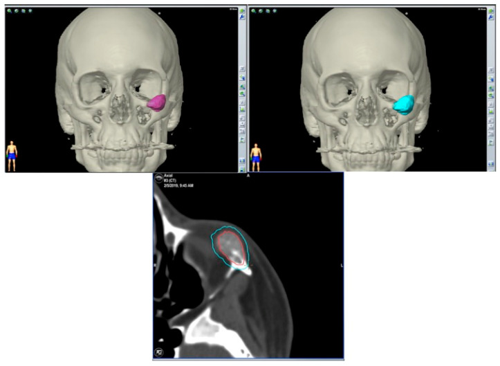
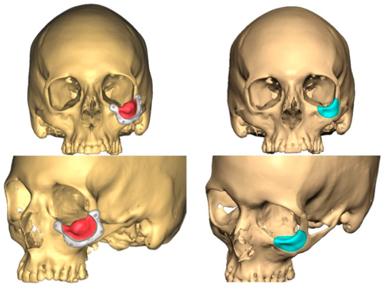
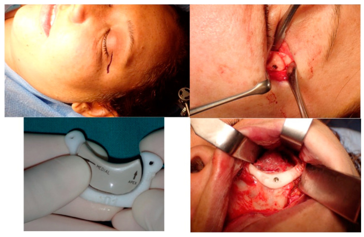
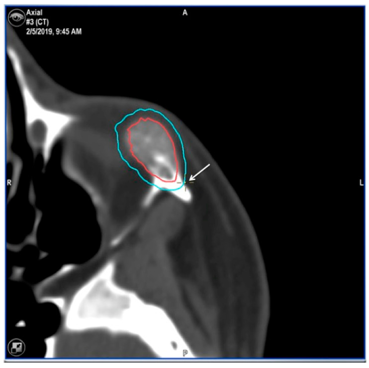
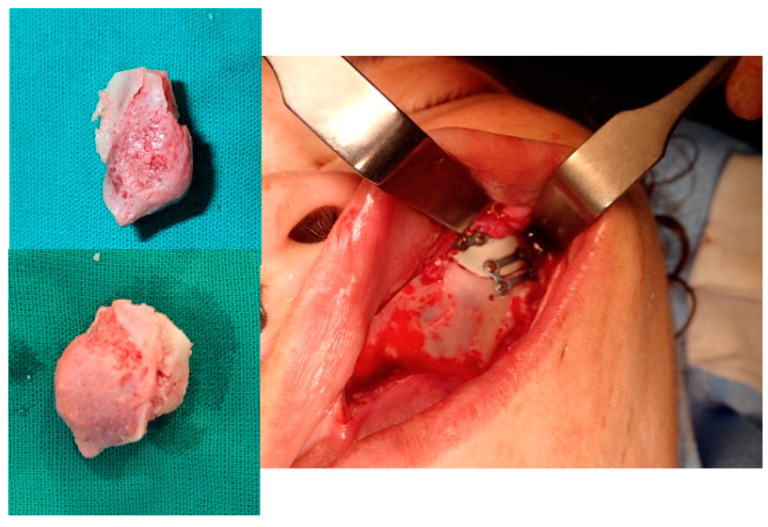
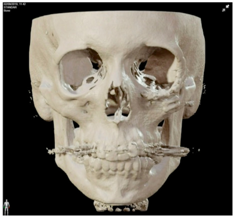
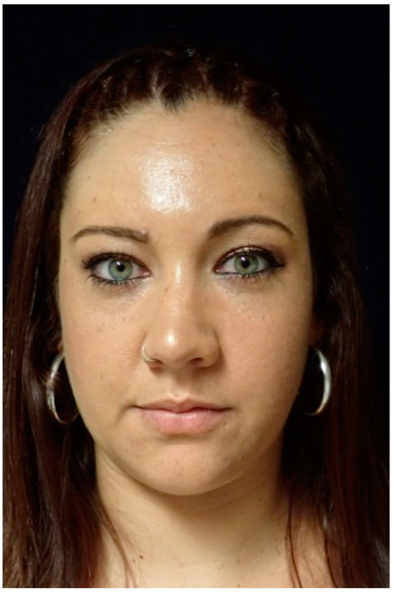
Background: Intraosseous vascular anomalies in the facial skeleton present significant diagnostic and therapeutic challenges due to complex anatomy. These anomalies represent about 0.5-1% of bony neoplastic and tumor-like lesions, usually presenting as a firm, painless mass. Most described intraosseous vascular malformations are venous malformations (VMs) and, more rarely, arteriovenous malformations. Objectives: The objectives of this work are to show our experience, protocol and the applications of computer planning, virtual surgery, CAD-CAM design, surgical navigation, and computer-assisted navigated piezoelectric surgery in the treatment of facial intraosseous vascular anomalies and to evaluate the advantages and disadvantages. Methods: Three females and one male with periorbital intraosseous vascular anomalies were treated using en-block resection and immediate reconstruction with a custom-made PEEK prosthesis. One lesion was in the supraorbital rim and orbital roof, one in the frontal bone and orbital roof, and two in the zygomatic region. We accomplished the resection and reconstruction of the lesion using virtual planning, CAD-CAM design, surgical navigation and piezoelectric device navigation. Results: There were no complications related to the surgery assisted with navigation. With an accuracy of less than 1 mm, the procedure may be carried out in accordance with the surgical plan. The surgeon's degree of uncertainty during deep osteotomies and in locations with low visibility was decreased by the use of the navigated piezoelectric device. Conclusions: Resection and reconstruction of facial intraosseous vascular anomalies benefit from this new surgical strategy using CAD-CAM technologies, computer-assisted navigated piezoelectric surgery, and surgical navigation.

Keywords: 3D planning; CAD-CAM design; computed-assisted surgery; computer-assisted navigated piezoelectric surgery; surgical navigation; virtual planning; virtual surgery.


Conflict of interest statement

The authors declare no conflicts of interest.

Figures

Figure 1
Figure 1
These images show the preoperative external appearance of the face of Patient 2. The protrusion can be seen in the right frontal region due to an intraosseous venous malformation.
Figure 2
Figure 2
Preoperative CT scan. Three-dimensional, axial, sagittal and coronal views can be appreciated. There is a mixed radiolucent lesion and an expansive lytic defect affecting the frontal bone above the right supraorbital rim and roof of the right orbit. A: anterior, P: posterior, H: head, F: foot, R: right, L: left.
Figure 3
Figure 3
The resection plan (in red in the figure) and the surgical cutting guide that will conduct the resection on the surface of the frontal bone as planned.
Figure 4
Figure 4
Navigated piezoelectric device. Direct or “live” navigation of the frontal bone. The skull post was anchored with a self-drilling screw.
Figure 5
Figure 5
Images of direct navigation with the piezoelectric device (yellow dot with small cross) are shown on the navigation screen. The progression and depth of the guided osteotomy can be appreciated and evaluated in real time (“live navigation”). A: anterior, P: posterior, H: head, F: foot, R: right, L: left. In blue, the virtual resection; in red, the virtual reconstruction with the PEEK prosthesis.
Figure 6
Figure 6
With the navigation pointer, the osteotomy (indirect navigation) we perform (green line on the navigation screen) can also be checked. A: anterior, P: posterior, H: head, F: foot, R: right, L: left. In blue, the virtual resection; in red, the virtual reconstruction with the PEEK prosthesis.
Figure 7
Figure 7
Images of direct navigation of the right orbital roof with the navigated piezoelectric device (yellow dot with small cross) are shown on the navigation screen. The progression and depth of the guided osteotomy can be appreciated and evaluated in real time (“live navigation”). A: anterior, P: posterior, H: head, F: foot, R: right, L: left. In blue, the virtual resection; in red, the virtual reconstruction with the PEEK prosthesis.
Figure 8
Figure 8
Postoperative CT scan. Three-dimensional, axial, sagittal and coronal views can be appreciated.
Figure 9
Figure 9
These images show the postoperative facial appearance with reestablishment of the frontal contour.
Figure 10
Figure 10
This image shows the preoperative external appearance of the face of Patient 3. There is a slight elevation of the left eyeball and a protrusion of the left cheek.
Figure 11
Figure 11
Preoperative CT scan. Three-dimensional, axial, sagittal and coronal views can be appreciated. There are a mixed radiolucent lesion and an expansive lytic defect affecting the left infraorbital rim, zygoma, external zone of the orbital floor and inferior part of the external wall.
Figure 12
Figure 12
Surgical plan with the BrainLab® navigation software, iPlan® 3.0.6 (Munich, Germany). The lesion is colored in red and the surgical margin in blue.
Figure 13
Figure 13
This image shows the lesion (in red), the surgical guide (in white) and the planned PEEK prosthesis (in blue).
Figure 14
Figure 14
This picture shows the surgical approach to the orbit, the surgical guide and the prosthesis, the vestibular intraoral approach with the surgical guide in position anchored to the zygoma with screws and the superior osteotomy line.
Figure 15
Figure 15
Images of direct navigation with the piezoelectric device on the less-visible posterior part the zygoma (yellow dot with small cross) are shown on the navigation screen. Again, the progression and depth of the guided osteotomy can be appreciated and evaluated in real time (“live navigation”). The lesion is colored in red and the surgical margin in blue.
Figure 16
Figure 16
The resected lesion, the PEEK prosthesis and the osteosynthesis with miniplates (intraoral approach).
Figure 17
Figure 17
Postoperative 3D CT.
Figure 18
Figure 18
Postoperative facial appearance.

Similar articles

References

    1. Liberale C., Rozell-Shannon L., Moneghini L., Nocini R., Tombris S., Colletti G. Stop calling me cavernous hemangioma! A literature review on misdiagnosed bony vascular anomalies. J. Investig. Surg. 2022;15:141–150. doi: 10.1080/08941939.2020.1824041. - DOI - PubMed
    1. Mulliken J.B., Glowacki J., Mulliken J.B., Glowacki J. Hemangiomas and vascular malformations in infants and children: A classification based on endothelial characteristics. Plast. Reconstr. Surg. 1982;69:412–422. doi: 10.1097/00006534-198203000-00002. - DOI - PubMed
    1. Behr G.G., Johnson C.J. Vascular anomalies: Hemangiomas and beyond- Part I, fast-flow lesions. Am. J. Roentgenol. AJR. 2013;200:414–422. doi: 10.2214/AJR.11.7852. - DOI - PubMed
    1. Wassef M., Blei F., Adams D., Alomari A., Baselga E., Berestein A., Burrows P., Frieden I.J., Garzon M.C., Lopez-Gutierrez J.C., et al. Vascular anomalies classification: Recommendations from the international society for the study of vascular anomalies. Pediatrics. 2015;136:e203–e214. doi: 10.1542/peds.2014-3673. - DOI - PubMed
    1. Kadlub N., Dainese L., Coulomb-L’Hermine A., Galmiche L., Soupre V., Ducou Lepointe H., Vazquez M.P., Picard A. Intraosseous haemangioma: Semantic and medical confusion. Int. J. Oral Maxillofac. Surg. 2015;44:718–724. doi: 10.1016/j.ijom.2015.01.025. - DOI - PubMed

LinkOut - more resources