. 2024 Aug 20;13(16):4903.
doi: 10.3390/jcm13164903.

Shear Wave Elastography of the Skin following Radial Forearm Free Flap Surgery in Transgender Patients: Observational Study

Affiliations

Shear Wave Elastography of the Skin following Radial Forearm Free Flap Surgery in Transgender Patients: Observational Study

Martino Guiotto et al. J Clin Med. .

Abstract

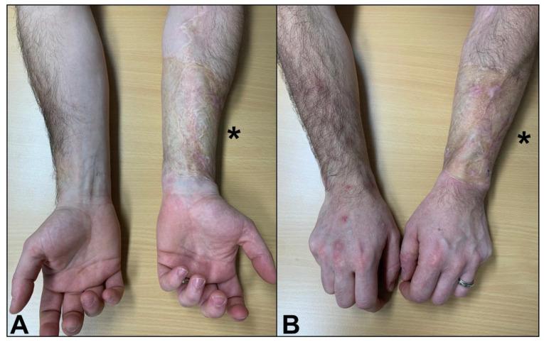
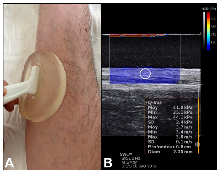
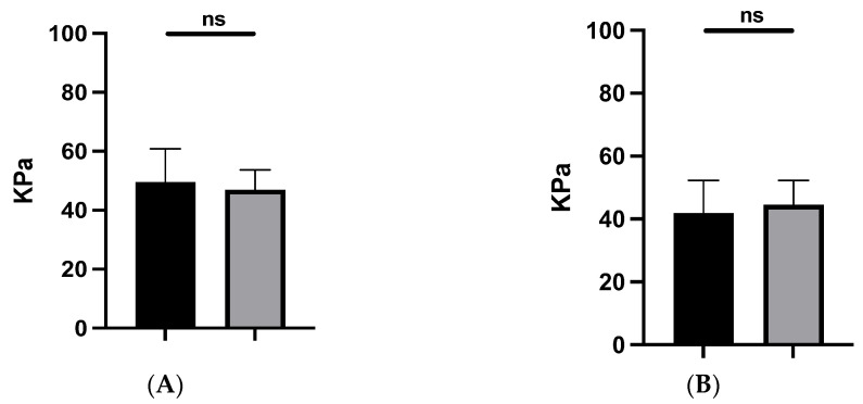
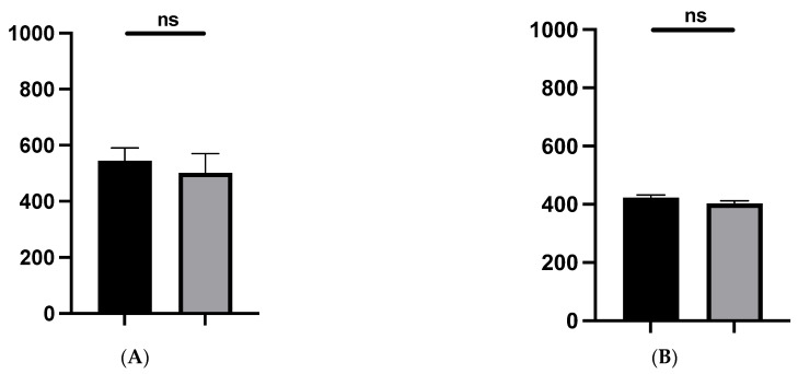
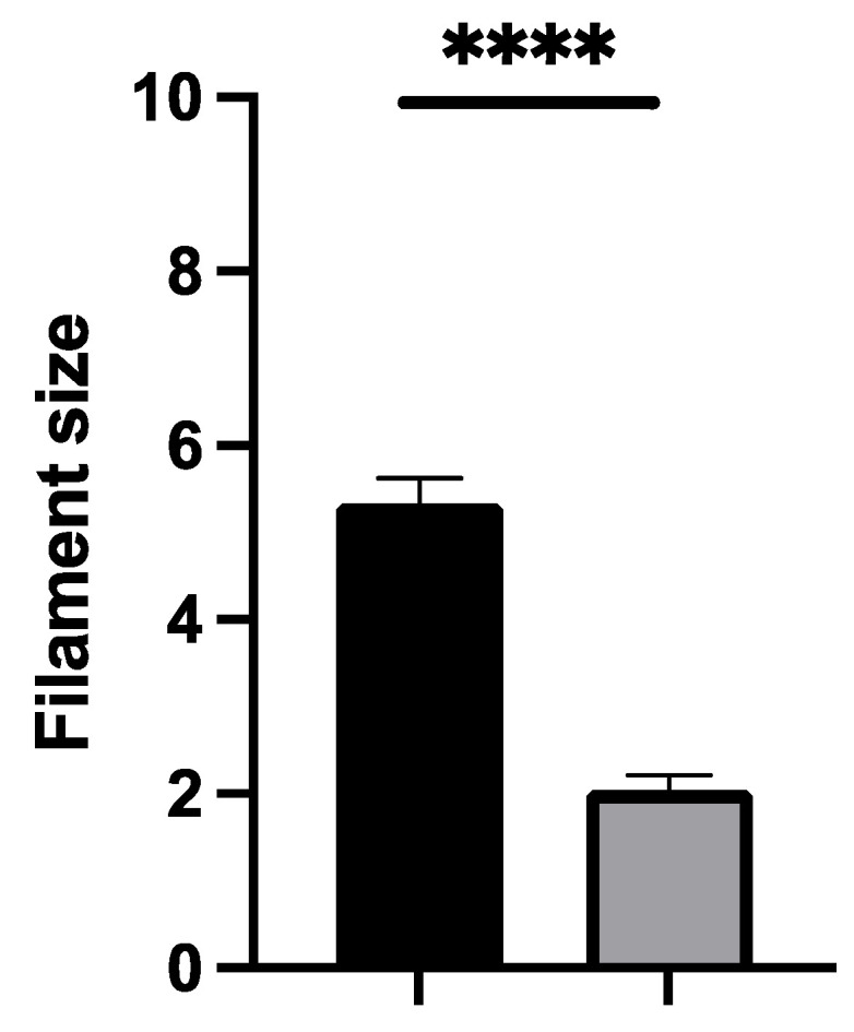
Background: Ultrasound shear wave elastography (SWE) noninvasively measures the stiffness of tissue by producing and measuring tissue deformation. Scar formation, a crucial aspect of wound healing, can lead to functional and aesthetic complications when pathological. While SWE has shown promise in dermatological evaluations, its role in surgical scar assessment remains underestimated. Our study aims to investigate SWE in evaluating surgical scars at the donor site after forearm free flap surgery in transgender patients. Methods: After radial forearm free flap harvesting, the donor site was grafted with a split-thickness skin graft with or without interposition of Matriderm. Eleven patients were evaluated more than one year after surgery, using SWE alongside scar characteristics, sensory outcomes, and patient satisfaction surveys. Results: Our study revealed no significant difference in stiffness (p > 0.15), pigmentation (p = 0.32), or erythema (p = 0.06) between operated and non-operated sides. The interposition of Matriderm did not influence the stiffness. Patients significantly (p < 0.0001) reported a loss of discrimination. Patients' subjective scar evaluation appeared in line with our quantitative and objective results. Conclusions: This study contributes to the evolving understanding of SWE's role in scar assessment, highlighting its feasibility in evaluating surgical scars. However, continued research efforts are necessary to establish SWE as a reliable and objective method for surgical scar evaluation and management.

Keywords: Patient and Observer Scar Assessment Scale (POSAS); radial forearm free flap; shear wave elastography; transgender; ultrasound; wound healing.


Conflict of interest statement

The authors declare no conflicts of interest.

Figures

Figure 1
Figure 1
Clinical outcome: anterior (A) and posterior (B) aspects of the skin-grafted donor site post-radial forearm free flap. *: donor site.
Figure 2
Figure 2
(A) Examination using a high-resolution 6–20 MHz linear array transducer with a thin-style gel pad. (B) The colored region represents the 2D quantitative elastogram superimposed on a B-mode image with a color scale (see top right). The software allowed us to measure the mean stiffness (Young’s modulus, in kPa) value and the shear wave velocity (m/s) of the skin inside a white circular region of interest (2 mm diameter Q-Box).
Figure 3
Figure 3
Shear wave modulus (KPa) of the skin in the transverse (A) and longitudinal (B) planes. Operated site (black) and contralateral site (grey). ns: not significant.
Figure 4
Figure 4
Pigmentation quantification (melanin) (A) and erythema (B) of the skin. Operated site (black) and contralateral site (grey). No notable difference in scar color, particularly in terms of inflammation (erythema) and melanin concentration, was observed between the operated (experimental) and non-operated (control) sides. ns: not significant.
Figure 5
Figure 5
Semmes–Weinstein monofilament test. Operated site (black) and contralateral site (grey). Patients reported a significant decrease in sensation (loss of discrimination) when tested using the Semmes–Weinstein monofilament test. This indicates impaired tactile sensitivity at the scar site compared to the non-operated side. ****: significant.

Similar articles

References

    1. Giambini H., An K.N. Ultrasound elastography for hand soft tissue assessment. Hand Clin. 2022;38:119–128. doi: 10.1016/j.hcl.2021.08.013. - DOI - PubMed
    1. Gennisson J.L., Deffieux T., Fink M., Tanter M. Ultrasound elastography: Principles and techniques. Diagn. Interv. Imaging. 2013;94:487–495. doi: 10.1016/j.diii.2013.01.022. - DOI - PubMed
    1. Sigrist R.M.S., Liau J., Kaffas A.E., Chammas M.C., Willmann J.K. Ultrasound elastography: Review of techniques and clinical applications. Theranostics. 2017;7:1303–1329. doi: 10.7150/thno.18650. - DOI - PMC - PubMed
    1. Lee S.Y., Cardones A.R., Doherty J., Nightingale K., Palmeri M. Preliminary Results on the Feasibility of Using ARFI/SWEI to Assess Cutaneous Sclerotic Diseases. Ultrasound Med. Biol. 2015;41:2806–2819. doi: 10.1016/j.ultrasmedbio.2015.06.007. - DOI - PMC - PubMed
    1. Billy J., Bensamoun S.F., Mercier J., Durand S. Applications of ultrasound elastography to hand and upper limb disorders. Hand Surg. Rehabil. 2024;43:101636. doi: 10.1016/j.hansur.2024.101636. - DOI - PubMed

LinkOut - more resources