Review
. 2024 Aug 7;25(16):8621.
doi: 10.3390/ijms25168621.

Divergent Processing of Cell Stress Signals as the Basis of Cancer Progression: Licensing NFκB on Chromatin

Affiliations
Review

Divergent Processing of Cell Stress Signals as the Basis of Cancer Progression: Licensing NFκB on Chromatin

Spiros A Vlahopoulos. Int J Mol Sci. .

Abstract

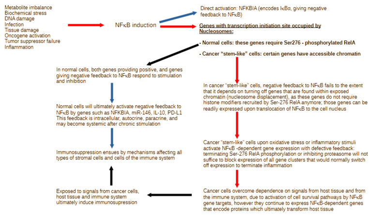
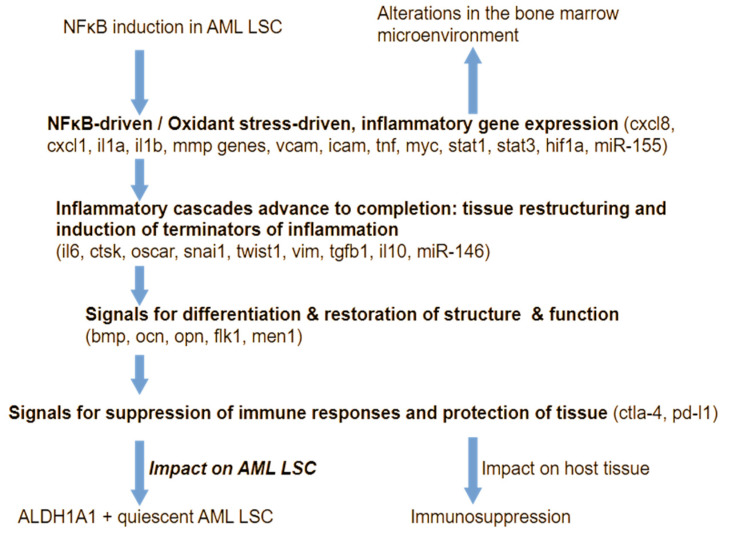
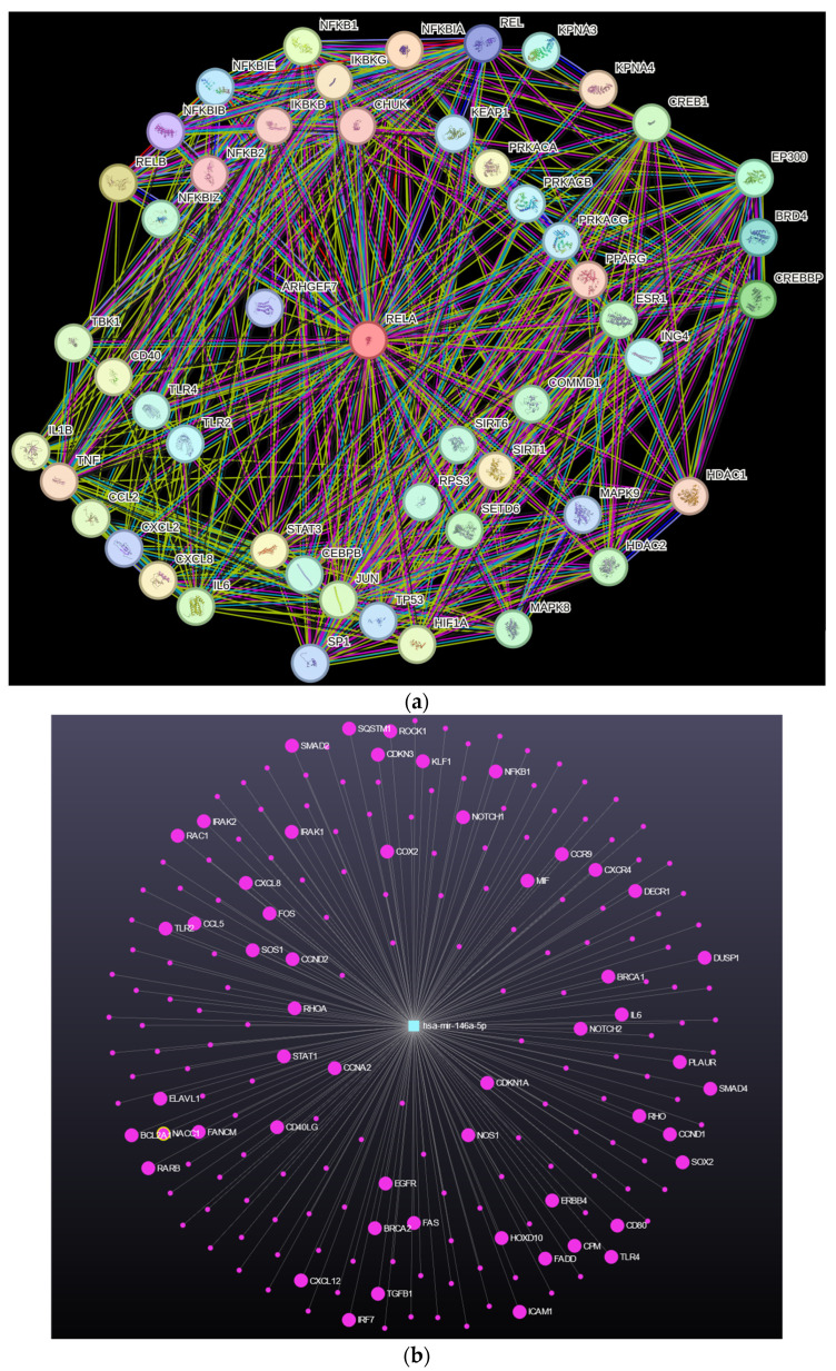
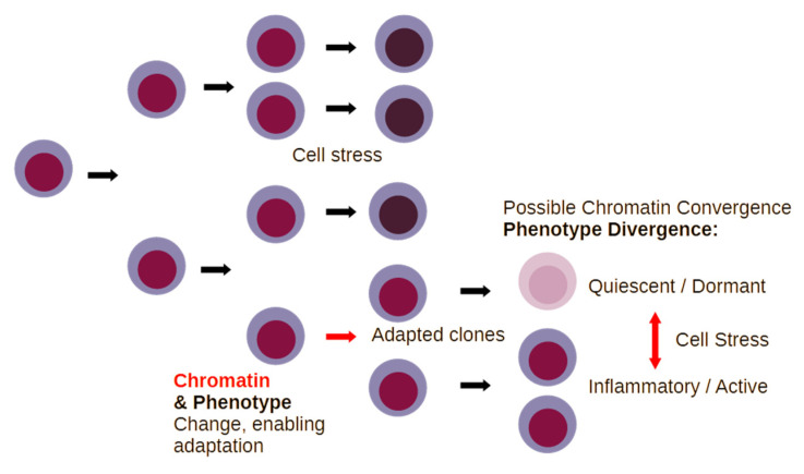
Inflammation is activated by diverse triggers that induce the expression of cytokines and adhesion molecules, which permit a succession of molecules and cells to deliver stimuli and functions that help the immune system clear the primary cause of tissue damage, whether this is an infection, a tumor, or a trauma. During inflammation, short-term changes in the expression and secretion of strong mediators of inflammation occur, while long-term changes occur to specific groups of cells. Long-term changes include cellular transdifferentiation for some types of cells that need to regenerate damaged tissue, as well as death for specific immune cells that can be detrimental to tissue integrity if they remain active beyond the boundaries of essential function. The transcriptional regulator NFκB enables some of the fundamental gene expression changes during inflammation, as well as during tissue development. During recurrence of malignant disease, cell stress-induced alterations enable the growth of cancer cell clones that are substantially resistant to therapeutic intervention and to the immune system. A number of those alterations occur due to significant defects in feedback signal cascades that control the activity of NFκB. Specifically, cell stress contributes to feedback defects as it overrides modules that otherwise control inflammation to protect host tissue. NFκB is involved in both the suppression and promotion of cancer, and the key distinctive feature that determines its net effect remains unclear. This paper aims to provide a clear answer to at least one aspect of this question, namely the mechanism that enables a divergent response of cancer cells to critical inflammatory stimuli and to cell stress in general.

Keywords: BET inhibitor; cell stress; chemokines; chromatin; cytokines; histone; inflammation; nuclear factor kappa B; super enhancer; unfolded protein response.


Conflict of interest statement

The author declares no conflict of interest.

Figures

Figure 1
Figure 1
In most cells, a variety of disruptions in tissue or cellular function activate NFκB posttranslational modifications and nuclear translocation, enabling cells to respond by expressing the genes that are needed to resolve the primary cause of stress. The NFKBIA (IκBα) gene is readily activated by NFκB and provides negative feedback. However, most other NFκB-driven genes (underlined bold text) require specific phosphorylation of Rel subunits (bold font), which recruit histone-remodeling complexes to increase chromatin accessibility (black arrows). The latter is not required for certain genes in the exposed chromatin of cancer “stem-like” cells. These cells are, therefore, permitted to express genes detrimental to the host tissue (red arrows). On the one hand, these cancer “stem-like” cells respond differently to cell stress and inflammation, and, on the other hand, their unrestricted expression of key modulators ultimately leads to changes in the host tissue and the immune system. The impact of such changes in gene regulation can have critical effects on tissue function. Blue arrows indicate expression and impact of genes that protect host tissue from excessive inflammation.
Figure 2
Figure 2
A simplified schematic of clone evolution for cancer cells under stress. Inflammation and cellular stress kill cancer cells by a number of different mechanisms. Defective responses to cell stress and inflammation, due to genetic or epigenetic inactivation of tumor suppressors under certain conditions, may permit the adaptation of cancer cells, which can give rise to either inflammatory or dormant cell clones. Specific aspects of chromatin accessibility, however, enable rather prompt switches between quiescent and inflammatory phenotypes in response to changes in tissue.
Figure 3
Figure 3
Inflammatory signal cascades generate conditions that restrict tumor growth, yet at the same time enable chromatin changes that permit phenotypic diversification and, consequently, the emergence of clones that are adapted to cell stress. Multiple different adapted clones, with diverse genetic assortments, may converge in the chromatin status, which is conducive to rapid changes in gene expression that permit adaptation to changing metabolic conditions and to altered interactions with the immune system. The type of malignant cell clones that are generated (red arrow) is critical to the severity of defects that these cells will cause to the host organism, ranging from paracrine effects to systemic defects in host immunity.
Figure 4
Figure 4
Gene expression is selectively unblocked in “stem-like” cells due to the accessible chromatin state, which permits the emergence of aggressive cancer (here AML) once a cell enters conditions that activate NFκB protein complexes. Cells that are not constitutively activating NFκB initially respond by positive feedback to inflammation, but ultimately activate negative feedback mechanisms that suppress inflammation and immunity, even if these cells are exposed to inflammatory stimuli. The nature of the cell signaling network architecture ensures redundancy in mechanisms for quenching inflammation. Cancer “stem-like” cells activate negative feedback to inflammation too, but incompletely, due to the perturbed functional state of their chromatin.
Figure 5
Figure 5
Illustrating the complexity of feedback mechanisms helps understand the depth of the impact of inflammation in cell signaling, which affects both conditions within the cell, as well as between the cell and its surrounding tissue. (a) A selection of “high-confidence” interacting proteins/genes with NFκB p65 (RelA) obtained with the platform string (https://string-db.org/, accessed on 6 July 2024). Details are clarified in the Supplementry Materials Figure S1. (b) An example of a key NFκB feedback gene is the micro RNA species miR146. This gene is prospectively associated with numerous other genes that influence cell stress responses (such as SQSTM1), inflammation (such as CXCL8), and cell phenotype (such as TGFβ1). Source miRNet 2.0 (https://www.mirnet.ca/, accessed on 6 July 2024).
Figure 6
Figure 6
A divergent response to cell stress characterizes malignant cells and shapes the pool of surviving malignant clones after changes in the conditions of their microenvironment. This phenomenon is especially evident in recurrent cancer. An additional reason why this is important lies in the fact that by proliferating, cancer cells gain the capacity to generate more phenotypically or genetically diversified clones (red arrows). By increasing clonal diversity, cancer becomes difficult to eradicate.

Similar articles

Cited by

References

    1. Lu H., Chen R., Barnie P.A., Tian Y., Zhang S., Xu H., Chakrabarti S., Su Z. Fibroblast Transdifferentiation Promotes Conversion of M1 Macrophages and Replenishment of Cardiac Resident Macrophages Following Cardiac Injury in Mice. Eur. J. Immunol. 2020;50:795–808. doi: 10.1002/eji.201948414. - DOI - PubMed
    1. Naitoh H., Suganuma Y., Ueda Y., Sato T., Hiramuki Y., Fujisawa-Sehara A., Taketani S., Araki M. Upregulation of Matrix Metalloproteinase Triggers Transdifferentiation of Retinal Pigmented Epithelial Cells in Xenopus Laevis: A Link between Inflammatory Response and Regeneration. Dev. Neurobiol. 2017;77:1086–1100. doi: 10.1002/dneu.22497. - DOI - PubMed
    1. Gaba A., Grivennikov S.I., Do M.V., Stumpo D.J., Blackshear P.J., Karin M. Cutting Edge: IL-10-Mediated Tristetraprolin Induction Is Part of a Feedback Loop That Controls Macrophage STAT3 Activation and Cytokine Production. J. Immunol. 2012;189:2089–2093. doi: 10.4049/jimmunol.1201126. - DOI - PMC - PubMed
    1. Stedile M., Lara Montero A., García Solá M.E., Goddio M.V., Beckerman I., Bogni E., Ayre M., Naguila Z., Coso O.A., Kordon E.C. Tristetraprolin Promotes Survival of Mammary Progenitor Cells by Restraining TNFα Levels. Front. Cell Dev. Biol. 2023;11:1265475. doi: 10.3389/fcell.2023.1265475. - DOI - PMC - PubMed
    1. Bao Y., Wu Y., Tao B., Sun R., Lin T., Zheng Y., Zhu X., Shen H., Chen W., Fan Y. Super-Enhancers Modulate Interleukin-6 Expression and Function in Cancers. Transl. Cancer Res. 2020;9:5555–5565. doi: 10.21037/tcr-19-2825. - DOI - PMC - PubMed

LinkOut - more resources