. 2024 Aug 9;25(16):8718.
doi: 10.3390/ijms25168718.

Lactoferrin Affects the Viability of Bacteria in a Biofilm and the Formation of a New Biofilm Cycle of Mannheimia haemolytica A2

Affiliations

Lactoferrin Affects the Viability of Bacteria in a Biofilm and the Formation of a New Biofilm Cycle of Mannheimia haemolytica A2

Lucero Ruiz-Mazón et al. Int J Mol Sci. .

Abstract

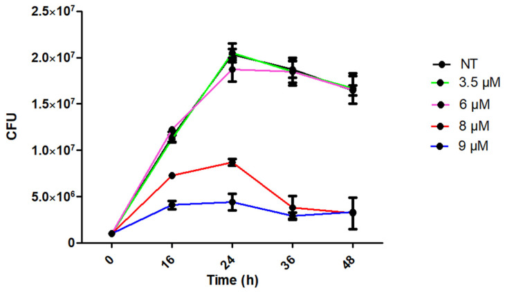
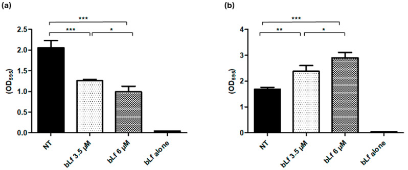
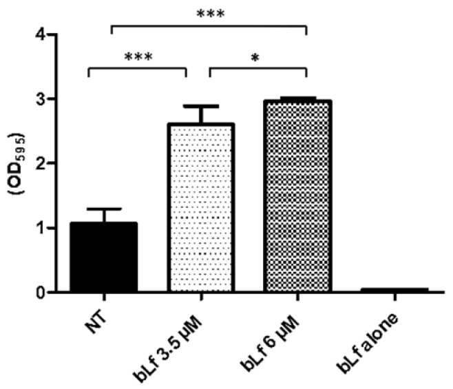
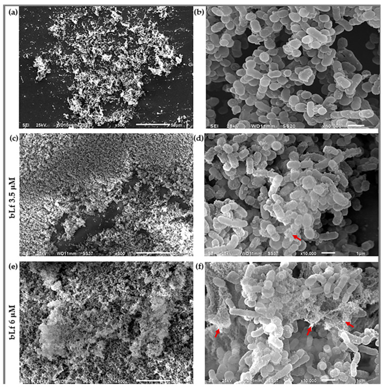
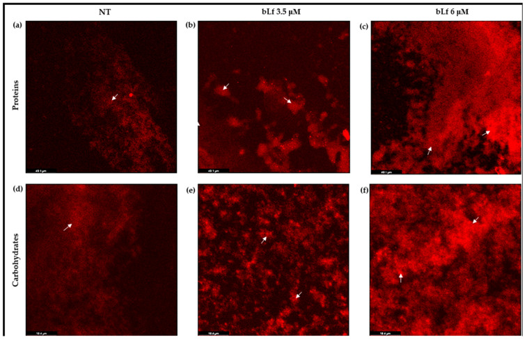
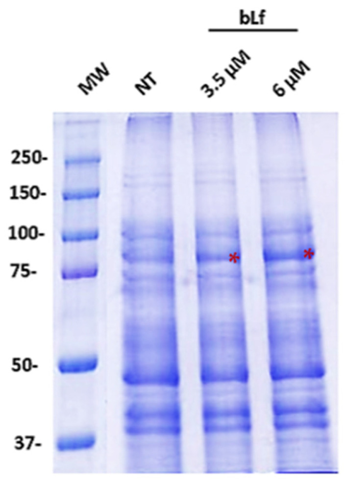
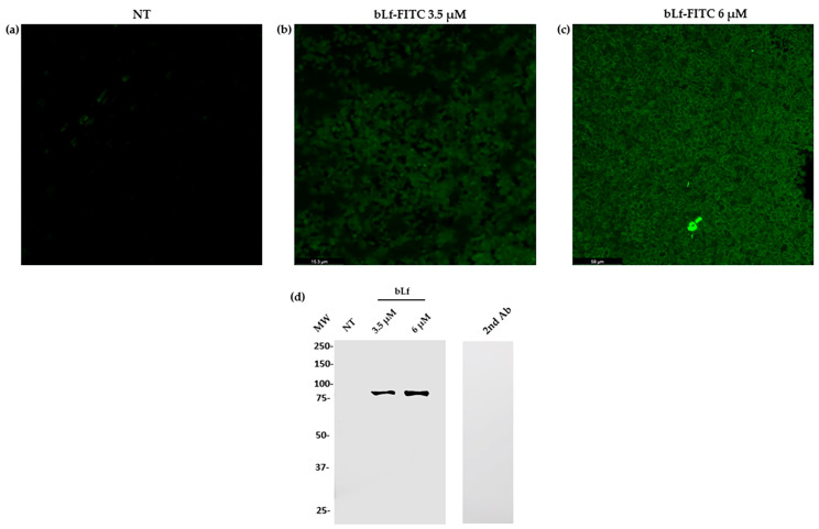
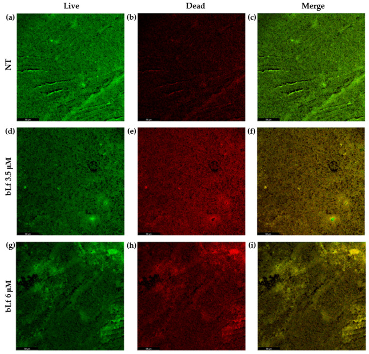
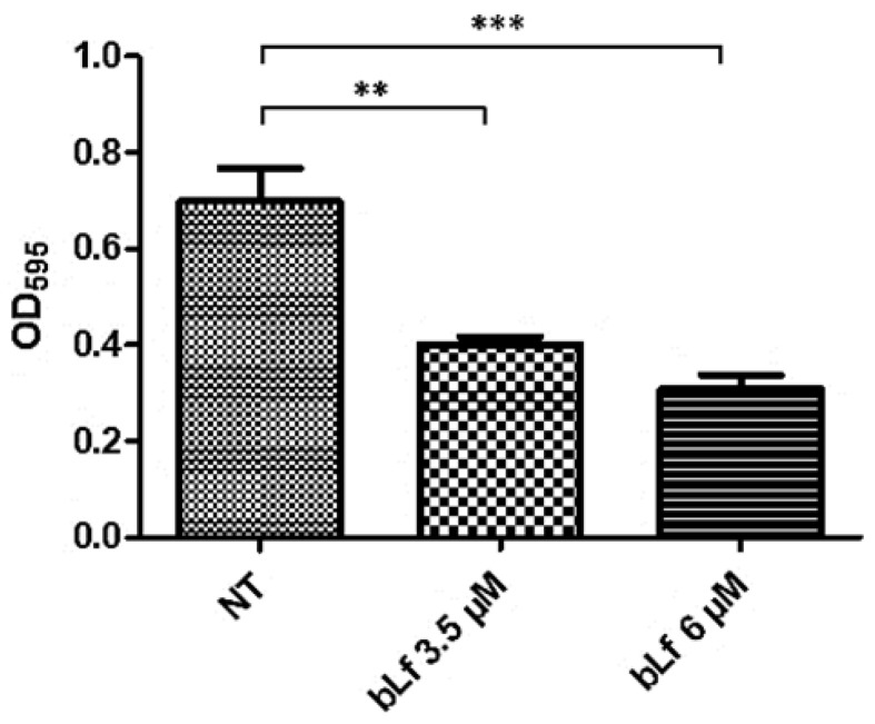
Respiratory diseases in ruminants are responsible for enormous economic losses for the dairy and meat industry. The main causative bacterial agent of pneumonia in ovine is Mannheimia haemolytica A2. Due to the impact of this disease, the effect of the antimicrobial protein, bovine lactoferrin (bLf), against virulence factors of this bacterium has been studied. However, its effect on biofilm formation has not been reported. In this work, we evaluated the effect on different stages of the biofilm. Our results reveal a decrease in biofilm formation when bacteria were pre-incubated with bLf. However, when bLf was added at the start of biofilm formation and on mature biofilm, an increase was observed, which was visualized by greater bacterial aggregation and secretion of biofilm matrix components. Additionally, through SDS-PAGE, a remarkable band of ~80 kDa was observed when bLf was added to biofilms. Therefore, the presence of bLf on the biofilm was determined through the Western blot and Microscopy techniques. Finally, by using Live/Dead staining, we observed that most of the bacteria in a biofilm with bLf were not viable. In addition, bLf affects the formation of a new biofilm cycle. In conclusion, bLf binds to the biofilm of M. haemolytica A2 and affects the viability of bacteria and the formation a new biofilm cycle.

Keywords: Mannheimia haemolytica; biofilm; bovine Lactoferrin; confocal laser microscopy.


Conflict of interest statement

The authors declare no conflicts of interest.

Figures

Figure 1
Figure 1
Viability of M. haemolytica A2 with bLf. M. haemolytica incubated with different concentrations of bLf at different times. The concentrations of 3.5 and 6 μM were sub-inhibitory until the 48th hour of incubation. Concentrations of 8 and 9 μM were inhibitory since they showed a significant difference regarding bacteria grown without bLf (NT) p < 0.05. Representative results of three independent experiments.
Figure 2
Figure 2
Inhibitory and stimulatory effect of bLf on the biofilm formation of M. haemolytica A2. (a) Bacteria were pre-incubated overnight with 3.5 and 6 μM bLf, then transferred to a microplate for biofilm formation; after 48 h, bacteria that had previous contact with bLf were no longer able to form a biofilm as bacteria without treatment do, since there was a reduction of 38 and 51% with incubation of 3.5 and 6 μM bLf, respectively. (b) Bacteria culture was transferred to a microplate for biofilm formation, and 3.5 and 6 μM bLf were added; after 48 h, an increase in biofilm was observed compared to the biofilm of untreated bacteria. These results were dependent on bLf concentration, as a significant difference was obtained between the different bLf concentrations used; bovine Lf alone was added to the microplate without biofilm to discard adherence of bLf to the microplate. Statistically significant differences between ratios are indicated (* p < 0.05, ** p < 0.01, *** p < 0.001). Representative results of three independent experiments.
Figure 3
Figure 3
Stimulatory effect of bLf on a mature biofilm of M. haemolytica A2. Bovine Lf (3.5 and 6 μM) was added to a 48 h biofilm of M. haemolytica A2. After 24 h, an increase in biofilm with bLf was observed, compared to a biofilm of bacteria with no treatment. These results were dependent on bLf concentration, as a significant difference was obtained between both bLf concentrations used, respectively; Lf alone was added to the microplate without biofilm to discard adherence of bLf to the microplate. Statistically significant differences between ratios are indicated (* p < 0.05, *** p < 0.001). Representative results of three independent experiments.
Figure 4
Figure 4
Biofilm observation under a Scanning Electron Microscope. Biofilm of M. haemolytica A2 with no treatment (NT) at a magnification of 500× (a) and 10,000× (b). The blank space shows the surface to which it is attached; biofilm with 3.5 μM bLf at a magnification of 500× (c) and 10,000× (d). The biofilm was observed to a greater extent and structures on the bacterial surface were noticed, compared to the biofilm with no treatment (red arrow). Biofilm with 6 μM bLf at a magnification of 500× (e) and 10,000× (f). A remarkable increase in biofilm was observed, indicated by the lack of blank space, as well as an increase in the structures on the bacterial surface and a mesh-like coating (red arrows). Representative images of three independent samples.
Figure 5
Figure 5
Proteins and carbohydrates of the biofilm matrix of M. haemolytica A2, observed under a laser confocal microscope. Images show proteins of the biofilm without bLf (NT) with the Sypro Ruby stain (ac), and carbohydrates of the biofilm with the Red Texas stain (df); proteins and carbohydrates from biofilm incubated with 3.5 μM bLf (b,e) and 6 μM bLf (c,f) are shown. The intensity of the fluorescence (white arrows) in the biofilms with bLf shows a major amount of these components compared to the biofilm with no treatment. Image magnification: 40×. Representative images of three independent samples.
Figure 6
Figure 6
Results of 10% SDS-PAGE of M. haemolytica A2 biofilm proteins incubated or unincubated with bLf. The protein pattern was analyzed by SDS-PAGE, using samples of 48 h biofilms with or without 3.5 and 6 μM bLf. With this assay, we did not observe differences in the biofilm pattern when bLf was added, in comparison to the biofilm without bLf. In addition, a remarkable band corresponding to the molecular weight of bLf (~80 kDa) was observed where it was added (red asterisks). Representative results of three independent samples.
Figure 7
Figure 7
Presence of bLf in the biofilm of M. haemolytica A2 shown through laser confocal microscopy and Western blot. Images show the distribution of bLf labeled with FITC (green fluorescence) at 3.5 μM (b) and 6 μM (c) throughout the biofilm of M. haemolytica A2, and a negative control where FITC-bLf was added to the plate with no biofilm (a). Western blot using anti-bLf in a biofilm without bLf (NT), with 3.5 or 6 μM bLf and a secondary anti-rabbit-HRP antibody alone as a negative control (d). Representative results of three independent samples.
Figure 8
Figure 8
Bovine Lf affects the viability of bacteria in biofilms with bLf, visualized by Confocal Laser Microscopy. CLM images of bacteria in biofilm for 48 h without bLf (NT) (ac) and with 3.5 (df) or 6 μM (gi) bLf, showing live bacteria with the Sypro stain (a,d,g) and dead bacteria with the propidium iodide exclusion stain (b,e,h). The merge shows a greater distribution of dead bacteria (visualized by the distribution of yellow fluorescence) in a biofilm with bLf (f,i) than in a biofilm without treatment, where a greater distribution of live bacteria was observed (visualized by the distribution of green fluorescence) (c). Representative images of three independent samples.
Figure 9
Figure 9
Formation of a new biofilm cycle of M. haemolytica A2. Bacteria of a 48 h biofilm with or without bLf were transferred to a new microplate to form a new biofilm cycle for another 48 h without bLf. A decrease in biofilm formation was observed in bacteria from a biofilm with bLf compared to bacteria from a biofilm with no treatment. Statistically significant differences between ratios are indicated (** p < 0.01, *** p < 0.001). Representative results of three independent experiments.

Similar articles

References

    1. Hussain R., Mahmood F., Ali H.M., Siddique A.B. Bacterial, PCR and Clinico-Pathological Diagnosis of Naturally Occurring Pneumonic Pasturellosis (Mannheimiosis) during Subtropical Climate in Sheep. Microb. Pathog. 2017;112:176–181. doi: 10.1016/j.micpath.2017.09.061. - DOI - PubMed
    1. Abdulkadir M., Nigussie T., Kebede I.A. Isolation and Identification of Pasteurella multocida and Mannheimia haemolytica from Pneumonic Small Ruminants and Their Antibiotic Susceptibility in Haramaya District, Eastern Ethiopia. Sci. World J. 2024;2024:5605552. doi: 10.1155/2024/5605552. - DOI - PMC - PubMed
    1. Singh K., Ritchey J.W., Confer A.W. Mannheimia haemolytica: Bacterial-Host Interactions in Bovine Pneumonia. Vet. Pathol. 2011;48:338–348. doi: 10.1177/0300985810377182. - DOI - PubMed
    1. Highlander S.K. Molecular Genetic Analysis of Virulence in Mannheimia (pasteurella) haemolytica. Front. Biosci. 2001;6:1128–1150. doi: 10.2741/highland. - DOI - PubMed
    1. het Lam J., Derkman T.H.J., van Garderen E., Dijkman R., van Engelen E. Distinct Mannheimia haemolytica Serotypes Isolated from Fatal Infections in Veal Calves and Dairy Cows. Vet. J. 2023;292:105940. doi: 10.1016/j.tvjl.2022.105940. - DOI - PubMed

LinkOut - more resources