. 2024 Aug 12;25(16):8779.
doi: 10.3390/ijms25168779.

Phase Separation of SARS-CoV-2 Nucleocapsid Protein with TDP-43 Is Dependent on C-Terminus Domains

Affiliations

Phase Separation of SARS-CoV-2 Nucleocapsid Protein with TDP-43 Is Dependent on C-Terminus Domains

Michael J Strong et al. Int J Mol Sci. .

Abstract

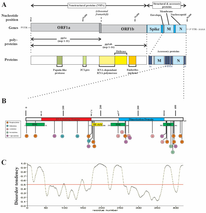
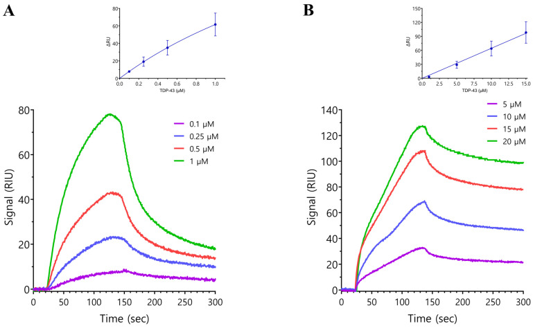
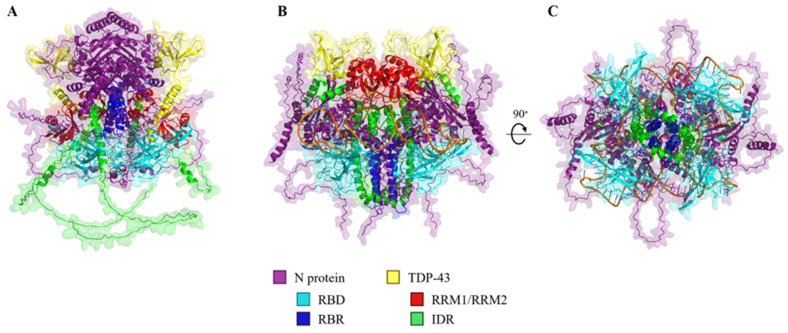
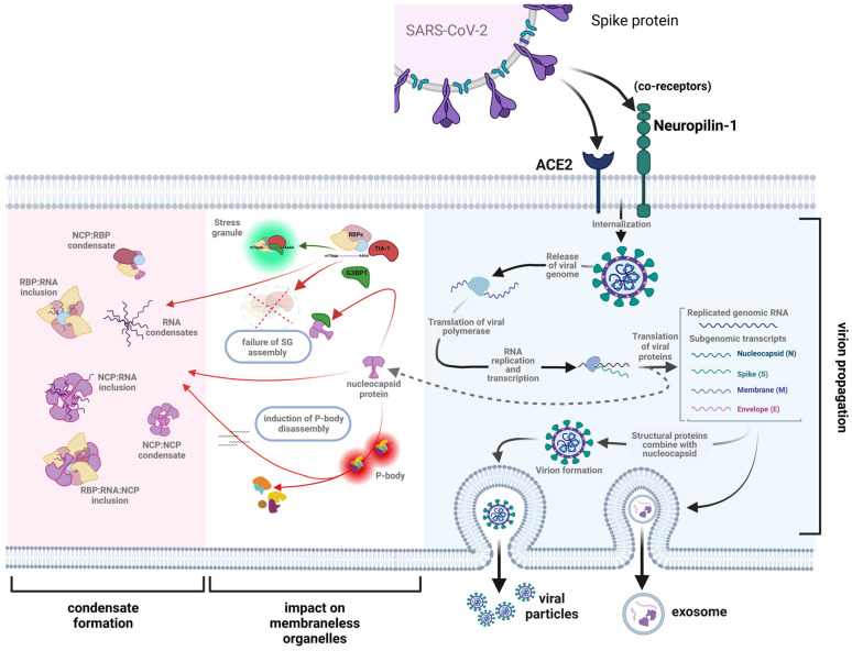
The SARS-CoV-2 nucleocapsid protein (N protein) is critical in viral replication by undergoing liquid-liquid phase separation to seed the formation of a ribonucleoprotein (RNP) complex to drive viral genomic RNA (gRNA) translation and in suppressing both stress granules and processing bodies, which is postulated to increase uncoated gRNA availability. The N protein can also form biomolecular condensates with a broad range of host endogenous proteins including RNA binding proteins (RBPs). Amongst these RBPs are proteins that are associated with pathological, neuronal, and glial cytoplasmic inclusions across several adult-onset neurodegenerative disorders, including TAR DNA binding protein 43 kDa (TDP-43) which forms pathological inclusions in over 95% of amyotrophic lateral sclerosis cases. In this study, we demonstrate that the N protein can form biomolecular condensates with TDP-43 and that this is dependent on the N protein C-terminus domain (N-CTD) and the intrinsically disordered C-terminus domain of TDP-43. This process is markedly accelerated in the presence of RNA. In silico modeling suggests that the biomolecular condensate that forms in the presence of RNA is composed of an N protein quadriplex in which the intrinsically disordered TDP-43 C terminus domain is incorporated.

Keywords: RNA binding proteins; amyotrophic lateral sclerosis; biomolecular condensates; neurodegeneration; nucleocapsid protein.


Conflict of interest statement

The authors declare no conflicts of interest.

Figures

Figure 1
Figure 1
A schematic representation of the SARS-CoV-2 nucleocapsid protein (N protein). (A) The structural protein segment of the SARS-CoV-2 gene encodes for multiple proteins, including the spike (S1 and S2 subunits), envelop (E), membrane (M), and nucleocapsid (N) proteins. (B) The N protein, encoded by the 9th ORF, is a 419 aa protein of 46 kDa composed of three domains: the N-terminus region contains a prominent RNA binding domain (RBD), a C-terminus dimerization domain that facilitates N protein dimer formation, and a Ser/Arg-rich central flexible linker region (LKR). The NCP undergoes extensive post-translational modification (PTM), predominantly within IDRs. Note the relative paucity of PTMs in the RBD—a feature not commonly observed in the RBPs of ALS-associated RBPs. Methylation at R95 is required for stress granule formation, while F17 has been shown to be the critical interactor for G3PB1 [23,27,28]. (C) The N protein contains three intrinsically disordered domains, including the N and C termini and the LKR (intrinsic disorder tendency predicted using PrDOS (protein disorder prediction system (https://prdos.hgc.jp/cgi-bin/result.cgi?ppid=387405p1d1696958772 (accessed on 10 October 2023)) and validated with UniProt (https://www.uniprot.org/uniprotkb/A0A2D1PX79) (accessed on 10 October 2023)). Modified from Strong, 2023 [1].
Figure 2
Figure 2
The relative aggregation of purified N and TDP-43 proteins in the absence of RNA (A), in the presence of polyrA (B), or in the presence of polyrG (C). Proteins and/or RNA were mixed at the same time in turbidity buffer with crowding reagent (10% dextran) and added to diluted Proteostat® aggregation detection reagent. The fluorescence shown was recorded with a plate reader approximately 30 min after initial mixing. N was mixed with the aggregate reagent at a high concentration (20–24 µM) or a low concentration (1–2.5 µM). N and full-length TDP-43 (flTDP-43) or N-terminus TDP-43 (TDP-431–269) were mixed at a molar ratio of 1:0.8. N was mixed with the RNA indicated at a molar ratio of 1:0.25. The same molar ratios were used in mixtures containing N, RNA, and TDP-43 variants. The fluorescence was normalized to the low N concentration (Nlow) used in each experiment. The data include on average three independent experiments with black dots showing these values.
Figure 3
Figure 3
Representative sensorgrams showing the interaction of FL-TDP-43 (A) or TDP-431–269 (B) with the immobilized SARS-CoV-2 N protein. Protein interactions were assessed by surface plasmon resonance using a Reichert 2SPR, SR7500DC System. The recombinant SARS-CoV-2 N protein was captured on the chip, TDP-43 analyte proteins were serially diluted in running buffer, and sensorgrams were determined at defined concentrations in at least three experiments. The equilibrium constant analysis using non-linear regression is shown with insets above the representative sensorgrams. The binding affinity (KD) values using steady-state analysis for the interaction with FL-TDP-43 and TDP-431–269 were 4.88 ± 1.13 μM and >167 μM, respectively, suggesting that the interaction between FL-TDP and the N protein is not mediated through the N-terminus domain of TDP-43.
Figure 4
Figure 4
In silico projections of the N protein: TDP-43 heteropolymer complex in the absence or presence of an RNA oligo. (A) Four N proteins are modeled in a complex with two FL-TDP-43 proteins. The N protein is displayed in purple, with the N-terminal RNA binding domain (RBD: residues 41–174) in cyan and the C-terminal RNA binding region (RBR: residues 210–246) in blue. TDP-43 is displayed in yellow, with the RRM1/RRM2 (residues 104–176 and 192–262) in red and the C-terminal intrinsically disordered region (IDR: residues 263–414) in green. (B) The same complex is modeled in the presence of two random 70-mer RNA oligos. (C) With a 90° rotation along the x-axis, this displays the bottom view of the heteropolymer. Note the more compact structure in which the IDR of TDP-43 becomes incorporated in proximity to the N-CTD.
Figure 5
Figure 5
A schematic illustration of the role of the SARS-CoV-2 nucleocapsid protein (N protein) in driving pathological biomolecular condensate formation. Upon endocytosis of the SARS-CoV-2 virus and release of the viral genome, the N protein plays a critical role in the translation of the viral gRNA and the formation of the virion. The latter is released either through exocytosis or encapsulated in exosomes. However, the free N protein also inhibits the formation of stress granules (SGs) by directly interacting with G3BP1, which is critical to the initial assembly of the SG. Not shown is the proposed additive role of the N protein in leading to the disassembly of processing bodies (PBs). The net effects are the formation of pathological biomolecular condensates of varying composition: N protein homopolymers; N protein: RNA heteropolymers; N protein: RBP heteropolymers; and N protein: RNA: RBP heteropolymers. It is hypothesized that any or all of these pathological molecular condensates would play a critical role in the pathogenesis of the neuronal or glial cytoplasmic inclusions that are the neuropathological hallmark of a broad array of neurodegenerative disorders (figure modified from Strong, 2023 [1] and created with BioRender.com).

Similar articles

Cited by

References

    1. Strong M.J. SARS-CoV-2, aging, and Post-COVID-19 neurodegeneration. J. Neurochem. 2023;165:115–130. doi: 10.1111/jnc.15736. - DOI - PMC - PubMed
    1. Pattanaik A., Bhandarkar B.S., Lodha L., Marate S. SARS-CoV-2 and the nervous system: Current perspectives. Arch. Virol. 2023;168:171. doi: 10.1007/s00705-023-05801-x. - DOI - PMC - PubMed
    1. Xu E., Xie Y., Al-Aly Z. Long-term neurologic outcomes of COVID-19. Nat. Med. 2022;28:2406–2415. doi: 10.1038/s41591-022-02001-z. - DOI - PMC - PubMed
    1. Hammarstrom P., Nystrom S. Viruses and amyloids—A vicious liaison. Prion. 2023;17:82–104. doi: 10.1080/19336896.2023.2194212. - DOI - PMC - PubMed
    1. Tsoi P.S., Quan M.D., Ferreon J.C., Ferreon A.C.M. Aggregation of Disordered Proteins Associated with Neurodegeneration. Int. J. Mol. Sci. 2023;24:3380. doi: 10.3390/ijms24043380. - DOI - PMC - PubMed

MeSH terms

LinkOut - more resources