Review
. 2024 Aug 13;25(16):8800.
doi: 10.3390/ijms25168800.

Ubiquitination Insight from Spinal Muscular Atrophy-From Pathogenesis to Therapy: A Muscle Perspective

Affiliations
Review

Ubiquitination Insight from Spinal Muscular Atrophy-From Pathogenesis to Therapy: A Muscle Perspective

Alfonso Bolado-Carrancio et al. Int J Mol Sci. .

Abstract

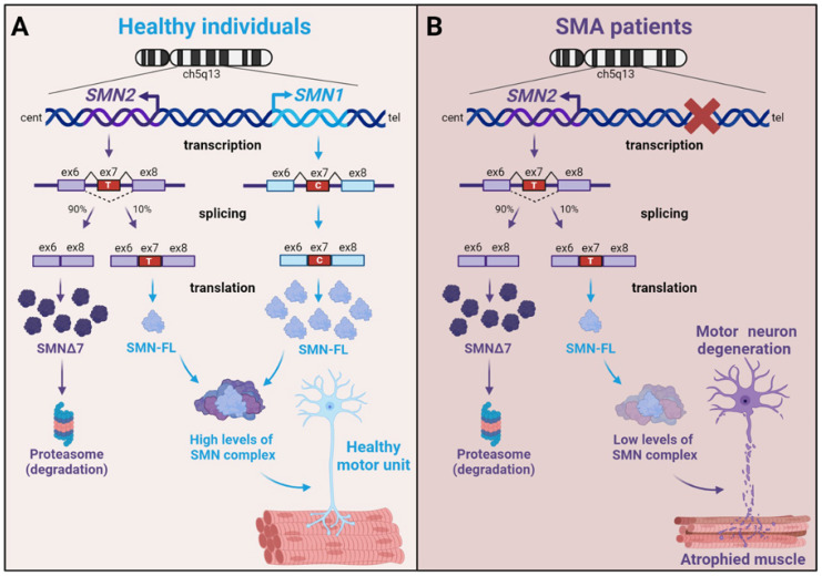
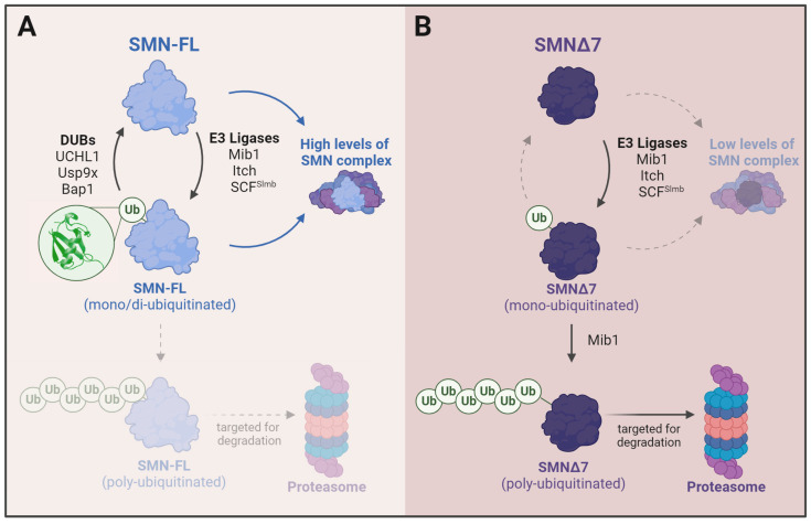
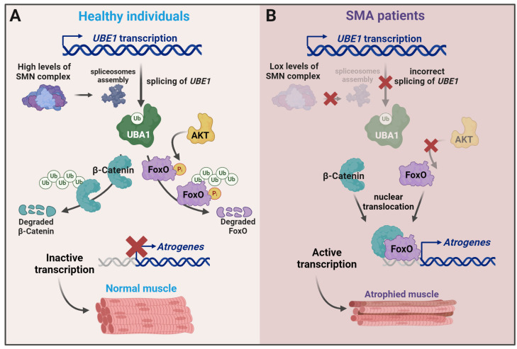
Spinal muscular atrophy (SMA) is one of the most frequent causes of death in childhood. The disease's molecular basis is deletion or mutations in the SMN1 gene, which produces reduced survival motor neuron protein (SMN) levels. As a result, there is spinal motor neuron degeneration and a large increase in muscle atrophy, in which the ubiquitin-proteasome system (UPS) plays a significant role. In humans, a paralogue of SMN1, SMN2 encodes the truncated protein SMNΔ7. Structural differences between SMN and SMNΔ7 affect the interaction of the proteins with UPS and decrease the stability of the truncated protein. SMN loss affects the general ubiquitination process by lowering the levels of UBA1, one of the main enzymes in the ubiquitination process. We discuss how SMN loss affects both SMN stability and the general ubiquitination process, and how the proteins involved in ubiquitination could be used as future targets for SMA treatment.

Keywords: SMN; skeletal muscle atrophy; spinal muscular atrophy; ubiquitin–proteasome system.


Conflict of interest statement

The authors declare no conflicts of interest.

Figures

Figure 1
Figure 1
Generation of SMN transcripts from SMN1 and SMN2 genes. (A) In healthy individuals, SMN1 and SMN2 gene transcripts are translated into the full-length SMN (SMN-FL) and SMNΔ7 proteins, respectively. A small percentage of SMN2 is also translated into SMN-FL. (B) In SMA patients, transcripts from SMN1 are absent. Most SMN2 transcripts are translated into SMNΔ7 and mostly degraded. See the text for a more detailed explanation. Figure created with BioRender.com.
Figure 2
Figure 2
Diagrammatic representation of the domain structure of SMN-FL. The SMN protein comprises several highly conserved motifs: a basic lysine-rich domain (K-rich), a Tudor domain, a poly-L-proline-rich domain (P-stretch), and a Y/G box in close proximity to the C-terminus, that itself mediates self-oligomerization and stability. The SMN protein is highly modified through phosphorylation (P), methylation (Me), acetylation (Ac), SUMOylation (S), and ubiquitination (Ub). Depicted are a summary of sites modified by the indicated PTMs identified through MS/proteomics and other methods (see [19] for a complete list of SMN PTMs sites). Some of the well-known proteins that interact with SMN-FL and the corresponding function are depicted below the corresponding interacting domain (see [20] for a review on SMN interactors and functional implications). Also indicated are the known interaction sites of SMN with the E3 UBLs Mib1, Itch and SCFSlmb and the DUB Bap1 (see Section 7 for a detailed discussion). Figure created with BioRender.com.
Figure 3
Figure 3
Outline of the ubiquitination process. An E1 ubiquitin-activating adenylates ubiquitin and forms an E1-Ub intermediate. Then, ubiquitin is transferred to the E2 ubiquitin-conjugating enzyme through a transthiolation reaction performed by E1. The E3 ligase forms an isopeptide bond between the substrate’s lysine side chain and the ubiquitin molecule’s C-terminal glycine. Monoubiquitinated molecules can either lose their ubiquitin moiety by the action of a deubiquitinase or become polyubiquitinated and further degraded in the proteasome. Figure created with BioRender.com.
Figure 4
Figure 4
(A) Mono-ubiquitination of SMN would not preclude its incorporation into the SMN complex. The incorporation of SMN into the complex, in turn, would prevent SMN from polyubiquitination and proteasomal degradation. Mono-ubiquitination can be reversed by Usp9x and Bap1. (B) The inability of SMNΔ7 to form stable complexes, its decreased affinity for Usp9x and its different compartment distribution would make SMNΔ7 more prone to polyubiquitination and degradation by the UPS. Figure created with Biorender.com.
Figure 5
Figure 5
A model to explain the functional link between SMN levels and ubiquitination and the changes in SMA. (A) Functional SMN complexes and correct UBE1 splicing render normal levels of UBA1 protein, which drive the ubiquitination and degradation of β-catenin and block the transcription of atrogenes. AKT activation reinforces the effect by phosphorylating FOXO and making it more sensitive to ubiquitination by MDM2. (B) In SMA patients, reduced levels of SMN result in low levels of UBA1, the accumulation of undegraded β-catenin, and increased atrogene expression. Figure created with Biorender.com.

Similar articles

References

    1. Perez-Baos S., Prieto-Potin I., Roman-Blas J.A., Sanchez-Pernaute O., Largo R., Herrero-Beaumont G. Mediators and Patterns of Muscle Loss in Chronic Systemic Inflammation. Front. Physiol. 2018;9:409. doi: 10.3389/fphys.2018.00409. - DOI - PMC - PubMed
    1. Pang X., Zhang P., Chen X., Liu W. Ubiquitin-proteasome pathway in skeletal muscle atrophy. Front. Physiol. 2023;14:1289537. doi: 10.3389/fphys.2023.1289537. - DOI - PMC - PubMed
    1. Ferreira R.P., Duarte J.A. Protein Turnover in Skeletal Muscle: Looking at Molecular Regulation towards an Active Lifestyle. Int. J. Sports Med. 2023;44:763–777. doi: 10.1055/a-2044-8277. - DOI - PubMed
    1. Murach K.A., Fry C.S., Dupont-Versteegden E.E., McCarthy J.J., Peterson C.A. Fusion and beyond: Satellite cell contributions to loading-induced skeletal muscle adaptation. FASEB J. 2021;35:e21893. doi: 10.1096/fj.202101096R. - DOI - PMC - PubMed
    1. Brzustowicz L.M., Lehner T., Castilla L.H., Penchaszadeh G.K., Wilhelmsen K.C., Daniels R., Davies K.E., Leppert M., Ziter F., Wood D., et al. Genetic mapping of chronic childhood-onset spinal muscular atrophy to chromosome 5q11.2-13.3. Nature. 1990;344:540–541. doi: 10.1038/344540a0. - DOI - PubMed

MeSH terms