. 2024 Aug 13;25(16):8802.
doi: 10.3390/ijms25168802.

Molecular Characteristics of the Malate Dehydrogenase (MDH) Gene Family in Spirometra mansoni (Cestoda: Diphyllobothriidea)

Affiliations

Molecular Characteristics of the Malate Dehydrogenase (MDH) Gene Family in Spirometra mansoni (Cestoda: Diphyllobothriidea)

Ruijie Wang et al. Int J Mol Sci. .

Abstract

The plerocercoid larva of Spirometra mansoni can cause a parasitic zoonosis-sparganosis. Malate dehydrogenase (MDH) plays a very important role in the life activities of parasites. However, little is known about the MDH family in S. mansoni. We identified eight new MDH members in S. mansoni in this study. Clustering analysis divided SmMDHs into two groups and revealed patterns similar to the conserved motif organization. RT-qPCR suggested that five MDHs were highly expressed in the mature proglottid and that three MDHs were highly expressed in the gravid proglottid. Phylogenetic analysis revealed that SmMDHs contain both conserved family members and members in the process of further diversification. rSmMDH has an NAD binding domain, a dimer interface and a substrate binding domain. Natural SmMDH was immunolocalized in the tissues and follicles around the uterus in the mature or gravid proglottid and eggshells. The maximum forward and reverse reaction activities of rSmMDH were observed at pH 8.5 and 9.0, respectively. The optimum temperature for enzyme activity was 37 °C in the forward reaction and 40 °C in the reverse reaction. These results lay the foundation for studying the molecular functions and mechanisms of MDHs in S. mansoni and related taxa.

Keywords: Spirometra mansoni; gene expression; malate dehydrogenase; molecular characterization; tapeworm.


Conflict of interest statement

The authors declare no conflict of interest.

Figures

Figure 1
Figure 1
Phylogenetic analysis of malate dehydrogenase in medical cestodes and trematodes based on the maximum likelihood method. The values on the branches represent bootstrap values, and only values with bootstrap values greater than 60 are presented.
Figure 2
Figure 2
Gene clustering and expression pattern analysis of S. mansoni. (a) Cluster analysis and conserved motifs of eight SmMDHs. According to the phylogenetic results, it can be divided into clade I and clade II. The numbers on the branches are bootstrap values, and only values above 60 are shown. (b) MDH gene expression of S. mansoni in different stages determined using qRT–PCR. Red represents the plerocercoid, blue represents the MP, green represents the GP, and orange represents the IMP. IMP: immature proglottid; MP: mature proglottid; GP: gravid proglottid. GAPDH was used as an internal reference gene. The expression level was measured with the 2-ΔΔCt method. The data were averaged from three repeats, The error bars represent the SDs (n = 3).
Figure 3
Figure 3
Molecular characterization of the cloned SmMDH. (a,b) The transcription pattern of the MDH gene in various developmental stages of S. mansoni including eggs, plerocercoids and adults. Conventional RT–PCR (a) and real-time RT–PCR (b) were performed. A housekeeping gene (GAPDH) was used as an internal reference. H2O was used as a negative control. IMP: immature proglottid; GP: gravid proglottid; MP: mature proglottid; Plero: plerocercoid. (c) soluble rSmMDH1 analysis. M: protein prestaining marker; Lane 1: uninduced bacterial cultures; Lane 2: the lysate of the induced recombinant bacteria harboring pQE-80L-rSmMDH1 after ultrasonication; Lane 3: the supernatant protein; Lane 4: the precipitate protein; (d) SDS–PAGE analysis after rSmMDH1 purification. M: protein prestaining marker; Lane 1: uninduced bacterial cultures; Lane 2: the lysate of the induced recombinant bacteria harboring pQE-80 L-rSmMDH1 after ultrasonication; Lane 3: rSmMDH1 purified by a Ni–NTA–sefinose column. (e) Determination of the optimal antigen coating concentration. (f) Anti-rSmMDH immunoserum potency assay. Red, blue, green, purple, yellow, and cyan represent serum dilutions of 1:102, 1:103, 1:104, 1:105, 1:106, and 1:107, respectively. (g) rSmMDH1 antigenicity analysis. M: Protein prestained marker; 1: rSmMDH1 + anti-rSmMDH1 serum; 2: rSmMDH1 + infected mouse serum; 3: rSmMDH1 + normal mouse serum; 4: soluble antigen + anti-rSmMDH1 serum; 5: soluble antigen + serum of infected mice; 6: soluble antigen + normal mouse serum; 7: ES antigen + anti-rSmMDH1 serum; 8: ES antigen + infected mouse serum; and 9: ES antigen + the serum of normal mice.
Figure 4
Figure 4
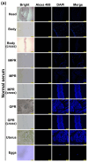
Immunofluorescence analysis of MDH in various stages of Spirometra mansoni. Head: head of plerocercoid; body: body of plerocercoid; body (cross): body of plerocercoid cross; MPR: mature proglottid; MPR (cross): mature proglottid cross; GPR: gravid proglottid; GPR (cross): gravid proglottid cross; IPR: immature proglottid; Egg: eggs in the uterus of gravid proglottid. (a) Normal serum; (b) infected serum; (c) anti-rSmMDH serum. GPR, scale of body (cross), head: 500 µm; IMPR, MPR, MPR (cross), GPR (cross), Uter, body: 200 µm; Eggs: 100 µm.
Figure 4
Figure 4
Immunofluorescence analysis of MDH in various stages of Spirometra mansoni. Head: head of plerocercoid; body: body of plerocercoid; body (cross): body of plerocercoid cross; MPR: mature proglottid; MPR (cross): mature proglottid cross; GPR: gravid proglottid; GPR (cross): gravid proglottid cross; IPR: immature proglottid; Egg: eggs in the uterus of gravid proglottid. (a) Normal serum; (b) infected serum; (c) anti-rSmMDH serum. GPR, scale of body (cross), head: 500 µm; IMPR, MPR, MPR (cross), GPR (cross), Uter, body: 200 µm; Eggs: 100 µm.
Figure 5
Figure 5
Enzymatic characteristics of SmMDH. (a) Kinetic study of the effects of the substrate concentration of NADH on the enzymatic activity of rSmMDH1. The kinetic parameters Km and Vmax were determined using Lineweaver–Burk plots. The Km and Vmax were 1.7507 mM and 0.9269 μmol min−1 mL−1, respectively. (b) Kinetic study of the effects of OAA substrate concentration on rSmMDH1 enzymatic activity. The Km and Vmax were 63.53 mM and 3.73 μmol min−1 mL−1, respectively. (c) Kinetic study of the effects of the malic acid concentration on the enzymatic activity of rSmMDH1. The Km and Vmax were 1.1933 mM and 0.071 μmol min−1 mL−1, respectively. (d) Kinetic study of the effects of the substrate concentration of NAD on the enzymatic activity of rSmMDH1. The Km and Vmax were 1.9489 mM and 0.098 μmol min−1 mL−1, respectively. (e) Effects of pH and temperature on rSmMDH1 enzymatic activity. (f) Effects of temperature on rSmMDH1 enzymatic activity. (g) Kinetic study effects of sodium dodecyl sulphate (SDS). (h) Kinetic study effects of thionicotinamide. (i) Kinetic study effects of (±) gossypol from cotton seeds.
Figure 6
Figure 6
Effects of different concentrations of thionicotinamide (Thio) on rSmMDH1 enzyme activity. Figure (a,c) show the oxalacetic acid oxidation of rSmMDH1. (a) The effect of different concentrations of thionicotinamide (Thio) on the initial velocities. (c) Thio (200, 600 µM). Figure (b,d) show the malic acid reduction of rSmMDH1. (b) The effect of different concentrations of thionicotinamide (Thio) on the initial velocities. (d) Thio (200, 600 µM). The inset shows a secondary plot of the 1/Vmax values derived from the primary Lineweaver–Burk plot vs. concentration for the determination of Ki.
Figure 7
Figure 7
Kinetic study of the effects of different compounds and metal ions. (a) Kinetics of nicotinic acid. (b) Kinetic study of the effects of an ethylenediaminetetraacetic acid disodium salt solution (EDTA-Na2). (c) Kinetic study of methanol. (d) Effect of different metal ions on the enzyme activity of the forward reaction. (e) Effect of different metal ions on the enzyme activity of the reverse reaction.

Similar articles

References

    1. Chen W.Q., Liu S.S., Cheng C., Cui J., Wang Z.Q., Zhang X. Molecular characteristics of glutathione transferase gene family in a neglect medical Spirometra tapeworm. Front. Vet. Sci. 2022;9:1035767. doi: 10.3389/fvets.2022.1035767. - DOI - PMC - PubMed
    1. Liu S., Gao F., Wang R., Li W., Wang S., Zhang X. Molecular Characteristics of the Fatty-Acid-Binding Protein (FABP) Family in Spirometra mansoni-A Neglected Medical Tapeworm. Animals. 2023;13:2855. doi: 10.3390/ani13182855. - DOI - PMC - PubMed
    1. Wang R.J., Li W., Liu S.N., Wang S.Y., Jiang P., Wang Z.Q., Zhang X. Integrated transcriptomic and proteomic analyses of plerocercoid and adult Spirometra mansoni reveal potential important pathways in the development of the medical tapeworm. Parasit Vectors. 2023;16:316. doi: 10.1186/s13071-023-05941-8. - DOI - PMC - PubMed
    1. Musrati R.A., Kollárová M., Mernik N., Mikulásová D. Malate dehydrogenase: Distribution, function and properties. Gen. Physiol. Biophys. 1998;17:193–210. - PubMed
    1. Madern D. Molecular evolution within the L-malate and L-lactate dehydrogenase super-family. J. Mol. Evol. 2002;54:825–840. doi: 10.1007/s00239-001-0088-8. - DOI - PubMed

LinkOut - more resources