. 2024 Aug 16;25(16):8909.
doi: 10.3390/ijms25168909.

Chikungunya and Mayaro Viruses Induce Chronic Skeletal Muscle Atrophy Triggered by Pro-Inflammatory and Oxidative Response

Affiliations

Chikungunya and Mayaro Viruses Induce Chronic Skeletal Muscle Atrophy Triggered by Pro-Inflammatory and Oxidative Response

Mariana Oliveira Lopes da Silva et al. Int J Mol Sci. .

Abstract

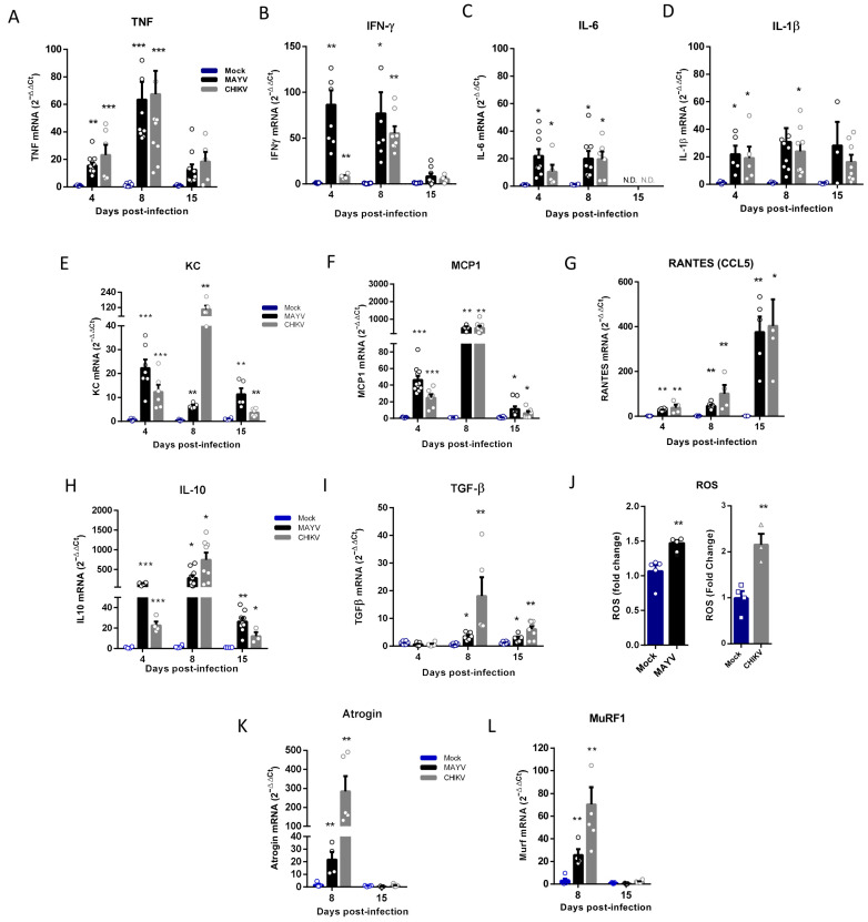
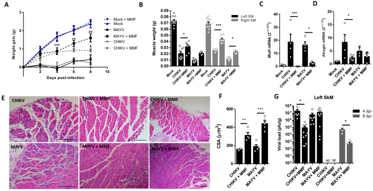
Chikungunya (CHIKV) and Mayaro (MAYV) viruses are arthritogenic alphaviruses that promote an incapacitating and long-lasting inflammatory muscle-articular disease. Despite studies pointing out the importance of skeletal muscle (SkM) in viral pathogenesis, the long-term consequences on its physiology and the mechanism of persistence of symptoms are still poorly understood. Combining molecular, morphological, nuclear magnetic resonance imaging, and histological analysis, we conduct a temporal investigation of CHIKV and MAYV replication in a wild-type mice model, focusing on the impact on SkM composition, structure, and repair in the acute and late phases of infection. We found that viral replication and induced inflammation promote a rapid loss of muscle mass and reduction in fiber cross-sectional area by upregulation of muscle-specific E3 ubiquitin ligases MuRF1 and Atrogin-1 expression, both key regulators of SkM fibers atrophy. Despite a reduction in inflammation and clearance of infectious viral particles, SkM atrophy persists until 30 days post-infection. The genomic CHIKV and MAYV RNAs were still detected in SkM in the late phase, along with the upregulation of chemokines and anti-inflammatory cytokine expression. In agreement with the involvement of inflammatory mediators on induced atrophy, the neutralization of TNF and a reduction in oxidative stress using monomethyl fumarate, an agonist of Nrf2, decreases atrogen expression and atrophic fibers while increasing weight gain in treated mice. These data indicate that arthritogenic alphavirus infection could chronically impact body SkM composition and also harm repair machinery, contributing to a better understanding of mechanisms of arthritogenic alphavirus pathogenesis and with a description of potentially new targets of therapeutic intervention.

Keywords: arthritogenic alphavirus; chronic atrophy; inflammation; oxidative stress; skeletal muscle.


Conflict of interest statement

The authors declare no conflicts of interest.

Figures

Figure 1
Figure 1
Temporal investigation of CHIKV and MAYV replication and clearance from SkM after subcutaneous infection in a young wild-type mice model. Wild-type (WT) SV129 mice of 12 day-olds were subcutaneously infected with MAYV or CHIKV in the left footpad, and tissues were collected at indicated time points. (A) Temporal quantification of viral load by plaque assay in the left gastrocnemius, (B) distribution in other tissues with detected infectious particles at 4 dpi and (C) at 8 dpi. (D) Virus- and Mock-infected mice swelling area of left paws and (E) weight gain was accompanied temporally. (F) Gastrocnemius muscle was dissected and immediately weighed at 4 dpi. Values were plotted as mean ± standard error of the mean (SEM). The inset shows a representative image of dissected muscles. Statistical analyses were performed to compare (AC) viral load of MAYV and CHIKV groups by multiple t-tests, and significance was determined using the Holm–Sidak method; (D,E) swelling and weight gain curve by two-way ANOVA and (F) muscle weight by one-way ANOVA followed by Tukey’s multiple comparison test from MAYV and CHIKV groups with mock. * p < 0.05, ** p < 0.01 and *** p < 0.001. Quad = quadriceps muscle. ND: not detected.
Figure 2
Figure 2
MAYV and CHIKV replication induces inflammatory infiltration and lesions, muscle mass loss, and fiber atrophy in skeletal muscle. (A) Left gastrocnemius of Mock or infected groups were collected at 4 dpi and stained with hematoxylin and eosin (H&E). Representative images from 2 infected mice demonstrate the inflammatory cell infiltration, fiber destruction, necrosis, and atrophic fibers in MAYV- and CHIKV-infected animals. High magnification images inset highlight the regions of atrophic fibers. Scale bars of figure = 100 μm and inset 50 μm. (B) Quantification of SkM fibers cross-sectional area (CSA) from H&E stained muscle images was performed using ImageJ 1.52a software. Each dot corresponds to the average CSA of fibers present in a field per mouse. (CE) The hind limbs of Mock- and MAYV-infected mice were imaged by NMRI. At each time point, images from five sections were acquired to calculate the area and then combined for volume reconstitution. (F) Expression levels of MuRF1 and Atrogin-1 in the left gastrocnemius at 4 dpi were determined by real-time PCR analysis. Cycle threshold (Ct) values were normalized to a housekeeping gene and analyzed using the ΔΔCt method to generate fold change values (2−ΔΔCt). Values are shown as mean ± standard error of the mean (SEM). Statistical analyses were performed using one-way ANOVA followed by Tukey’s multiple comparison tests (B,F), and for the comparison of tissue volumes of Mock and MAYV groups, multiple t-tests were used, and the significance was determined by the Holm–Sidak method. * p < 0.05, ** p < 0.01 and *** p < 0.001. Image (C) was created with BioRender.com.
Figure 3
Figure 3
Skeletal muscle atrophy and genomic RNA persist in the late phase of MAYV and CHIKV infection. Wild-type (WT) SV129 mice of 12-day-olds were infected, and SkMs were analyzed at late times post-infection. (A) Body weight was recorded until 30 dpi, and (B) gastrocnemius muscles were dissected and immediately weighed. (C) CHIKV and MAYV RNA were detected by TaqMan real-time PCR analysis. The dotted lines represent the Ct value limit for positive samples. (D) Temporal histological analyses of left gastrocnemius stained with H&E and respective quantification of SkM fiber cross-sectional area (CSA) from H&E stained muscle images using ImageJ 1.52a software. Each dot corresponds to the average CSA of fibers present in a field per mouse. Scale bars of figure = 100 μm and inset 50 μm. Values are shown as mean ± standard error of the mean (SEM). Statistical analysis of the weight gain curve comparing MAYV and CHIKV groups with Mock was performed by two-way ANOVA (A); for muscle weight (B) and CSA (D), one-way ANOVA was performed, followed by Tukey’s multiple comparison test. * p < 0.05, ** p < 0.01 and *** p < 0.001 for Mock and MAYV comparison; # p < 0.05, ## p < 0.01 and ### p < 0.001 for Mock and CHIKV comparison.
Figure 4
Figure 4
Temporal analysis of inflammatory mediator and atrogen expression in the early and late phases of MAYV and CHIKV infection. Wild-type (WT) SV129 mice of 12 day-olds were subcutaneously infected with MAYV or CHIKV in the left footpad, and left gastrocnemius were collected at indicated time points. (AI,K,L) Quantification of gene expression using real-time PCR analysis. Cycle threshold (Ct) values were normalized to a housekeeping gene and analyzed using the ΔΔCt method to generate fold change values (2−ΔΔCt). (J) Total reactive oxygen species (ROS) production in the left gastrocnemius was determined by fluorescence analysis using DCFDA. Arbitrary values of fluorescence from each sample were obtained at the end of the DCF incubation and plotted as fold change from the media of mock values. Values were plotted as mean ± standard error of the mean (SEM). Statistical analyses were performed by multiple t-tests to compare Mock with CHIKV and MAYV groups, and the significance was determined using the Holm–Sidak method. * p < 0.05, ** p < 0.01, and *** p < 0.001.
Figure 5
Figure 5
Treatment with Inflixmab reduces muscle mass loss and atrogen expression induced by CHIKV infection. Wild-type (WT) SV129 mice of 12 day-olds were subcutaneously infected with CHIKV and then treated with a daily dose of 20 μg of Infliximab (IFX) or Vehicle (PBS) by i.p. inoculation. (A) Weight gain was accompanied daily, and (B) gastrocnemius was dissected at 4 dpi for muscle weight measurement, (C) Atrogin expression by real-time PCR, and (D) viral load by plaque assay. (E) Virus- and Mock-infected mice swelling area of left paws was measured daily. Values were plotted as mean ± standard error of the mean (SEM). Statistical analyses of weight gain curve and paw swelling comparing treated and untreated CHIKV infected groups were performed by two-way ANOVA (A,E); and for muscle weight (B), Atrogin-1 expression (C), and viral load (D), by one-way ANOVA followed by Tukey’s multiple comparison test. * p < 0.05, ** p < 0.01 and *** p < 0.001.
Figure 6
Figure 6
Treatment with MMF reduces muscle atrophy and inflammation induced by MAYV and CHIKV infection. Wild-type (WT) SV129 mice of 12 day-olds were intraperitoneally treated with 20 mg/kg of monomethyl fumarate (MMF) or Vehicle (DMSO) and after 4 h infected with MAYV or CHIKV in the left footpad. (A) Weight gain was accompanied daily. Gastrocnemius were dissected at 8 dpi for analysis of (B) muscle weight, (C) MuRF1, and (D) Atrogin-1 expression by real-time PCR. (E) Histological analysis of left gastrocnemius stained with H&E (Scale bars of figures = 100 μm) and (F) respective quantification of SkM fiber cross-sectional area (CSA) from muscle images using ImageJ 1.52a software. (G) Viral load at SkM was determined by plaque assay at 4 and 8 dpi. Values were plotted as mean ± standard error of the mean (SEM). ND: not detected. Statistical analyses of weight gain curve comparing treated and untreated CHIKV and MAYV infected groups were performed by two-way ANOVA (A) with * used for indicates significance of comparison of both MAYV and CHIKV groups (4 and 6 dpi); # Only MAYV, and & CHIKV groups (8 dpi); one-way ANOVA was used in the analysis of muscle weight (B), Atrogens expression (C,D), CSA (F), and viral load (G), followed by Tukey’s multiple comparison test. */& p < 0.05, **/## p < 0.01 and *** p < 0.001.
Figure 7
Figure 7
Schematic representation of the possible mechanism involved in arthritogenic alphavirus-induced early and late skeletal muscle atrophy. ① At physiological conditions, young mice SkM fiber growth throughout life is determined by individual muscle fiber enlargement and mass gain by a higher rate of synthesis than protein degradation. ② New fiber generation by myogenesis will be recruited mainly after injury by activation of quiescent satellite cells. ③–⑧ Arthritogenic alphavirus replicates in SkM fibers, resulting in fiber destruction, recruitment of immune cells, and the production of inflammatory mediators in the early phase of infection. ⑨–⑩ Inflammation-induced protein degradation by UPS through activation of MuRF1 and Atrogin-1 that drives acute SkM atrophy. ⑪–⑫ Treatment with IFX or MMF reduces protein degradation and atrophy, corroborating the involvement of these pathways in MAYV- and CHIKV-induced atrophy. ⑬ SkM atrophy persists in the late phase of infection. ⑭ Despite the decrease in TNF, IFNγ, and IL-6 and also atrogen expression levels, ⑮–⑰ chemokines, IL-10, TGFβ mediators, and viral genomic RNA are still detected, indicating an incomplete viral clearance. ⑱ Long-term immune activation results in a reduction in CSA by a UPS-independent mechanism. Symbols indicates: formula image block/inhibition; formula image positive regulation; formula image open ended questions.

Similar articles

References

    1. Suhrbier A., Jaffar-Bandjee M.C., Gasque P. Arthritogenic alphaviruses—An overview. Nat. Rev. Rheumatol. 2012;8:420–429. doi: 10.1038/nrrheum.2012.64. - DOI - PubMed
    1. Santiago F.W., Halsey E.S., Siles C., Vilcarromero S., Guevara C., Silvas J.A., Ramal C., Ampuero J.S., Aguilar P.V. Long-Term Arthralgia after Mayaro Virus Infection Correlates with Sustained Pro-inflammatory Cytokine Response. PLoS Negl. Trop. Dis. 2015;9:e0004104. doi: 10.1371/journal.pntd.0004104. - DOI - PMC - PubMed
    1. Poidinger M., Scherret J.H., Broom A.K., Turley P.J., Lindsay M.D., Roy S., Mackenzie J.S., Hall R.A. Genetic stability among temporally and geographically diverse isolates of Barmah Forest virus. Am. J. Trop. Med. Hyg. 1997;57:230–234. doi: 10.4269/ajtmh.1997.57.230. - DOI - PubMed
    1. Tesh R.B., Watts D.M., Russell K.L., Damodaran C., Calampa C., Cabezas C., Ramirez G., Vasquez B., Hayes C.G., Rossi C.A., et al. Mayaro virus disease: An emerging mosquito-borne zoonosis in tropical South America. Clin. Infect. Dis. 1999;28:67–73. doi: 10.1086/515070. - DOI - PubMed
    1. Taylor S.F., Patel P.R., Herold T.J. Recurrent arthralgias in a patient with previous Mayaro fever infection. South Med. J. 2005;98:484–485. doi: 10.1097/01.SMJ.0000145879.14102.F4. - DOI - PubMed

MeSH terms

LinkOut - more resources