Comparative Study
. 2024 Aug 16;25(16):8926.
doi: 10.3390/ijms25168926.

The Comparison of Immunomodulatory Properties of Canine and Human Wharton Jelly-Derived Mesenchymal Stromal Cells

Affiliations
Comparative Study

The Comparison of Immunomodulatory Properties of Canine and Human Wharton Jelly-Derived Mesenchymal Stromal Cells

Anna Burdzinska et al. Int J Mol Sci. .

Abstract

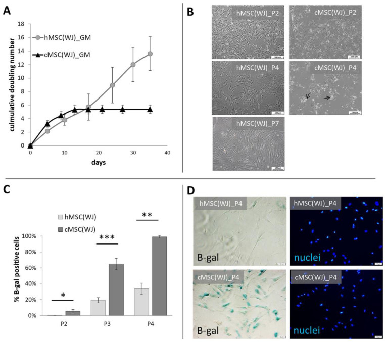
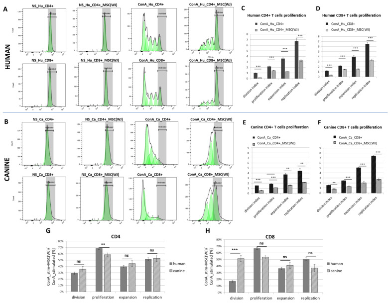
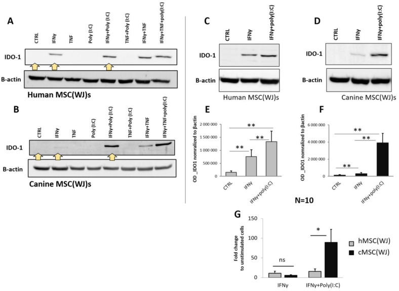
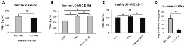
Although therapies based on mesenchymal stromal cells (MSCs) are being implemented in clinical settings, many aspects regarding these procedures require further optimization. Domestic dogs suffer from numerous immune-mediated diseases similar to those found in humans. This study aimed to assess the immunomodulatory activity of canine (c) Wharton jelly (WJ)-derived MSCs and refer them to human (h) MSCs isolated from the same tissue. Canine MSC(WJ)s appeared to be more prone to in vitro aging than their human counterparts. Both canine and human MSC(WJ)s significantly inhibited the activation as well as proliferation of CD4+ and CD8+ T cells. The treatment with IFNγ significantly upregulated indoleamine-2,3-dioxygenase 1 (IDO1) synthesis in human and canine MSC(WJ)s, and the addition of poly(I:C), TLR3 ligand, synergized this effect in cells from both species. Unstimulated human and canine MSC(WJ)s released TGFβ at the same level (p > 0.05). IFNγ significantly increased the secretion of TGFβ in cells from both species (p < 0.05); however, this response was significantly stronger in human cells than in canine cells. Although the properties of canine and human MSC(WJ)s differ in detail, cells from both species inhibit the proliferation of activated T cells to a very similar degree and respond to pro-inflammatory stimulation by enhancing their anti-inflammatory activity. These results suggest that testing MSC transplantation in naturally occurring immune-mediated diseases in dogs may have high translational value for human clinical trials.

Keywords: canine mesenchymal stromal cells; human mesenchymal stromal cells; immunomodulation; indoleamine 2,3-dioxygenase 1; transforming growth factor beta; umbilical cord.


Conflict of interest statement

The authors declare no conflicts of interest.

Figures

Figure 1
Figure 1
Growth dynamics and senescence of human and canine umbilical cord-derived MSCs in standard conditions. (A) Cumulative doubling number of representative human (h) and canine (cMSC(WJ)) cells which were cultured for 35 days (day 0—1st passage), (mean values, SD, n = 3). (B) The appearance of human (left column) and canine (right column) cells in standard conditions after the 2nd (P2, upper row),4th (P4, middle row), and 7th (P7, lower picture, human only) passage. Black arrows indicate fragmented cells, scale bars–100 μm (C) The mean (±SEM) proportion of cells displaying senescence associated β-galactosidase (β-gal) activity in human and canine populations on subsequent passages (p). *, p < 0.05; **, p < 0.01; ***, p < 0.001, Student’s t-test, n = 5. (D) Assessment of cell senescence using microscopy: images in rows present the same fields of view. Left column—cells with β-gal activity are blue, right column—cell nuclei stained in blue, scale bars–50 μm.
Figure 2
Figure 2
The effect of different culture conditions on population doubling time (PDT) and senescence of canine Wharton jelly-derived mesenchymal stromal cells, MSC(WJ). (A) PDT of canine MSC(WJ) cultured in growth medium (GM), GM with 2.5 ng/mL FGF2 (FGF2), in GM on gelatin-coated surface (GEL), and GM + FGF2 on gelatin-coated surface (FGF2_GEL), n = 6, *—p < 0.05 in comparison to control using Wilcoxon test or Student’s t-test for related data test depending on data distribution; (B) The proportion of senescent cells within the population (mean ±SEM) cultured in GM, FGF, and FGF2_GEL in subsequent passages (p). Data analyzed within each passage using one-way ANOVA with post hoc Tukey’s test. *, p < 0.05; **, p < 0.01; ***, p < 0.001; n ≥ 5; (C) representative images used for calculation β-gal assay (using X-gal substrate for reaction). Images in columns represent the same field of view. Upper row—light microscopy, senescent cells visible as blue, lower row—fluorescent microscopy, cell nuclei stained in red. Scale bars—50 μm. (D) The comparison of cell senescence at third passage (P3) between human MSC(WJ)s cultured in GM on uncoated surface and canine MSC(WJ) cultured in FGF2_GEL conditions. The graph presents median, quartiles, min and max values, n = 8. Data analyzed using Mann–Whitney U assay. ns—p > 0.05.
Figure 3
Figure 3
Human Wharton jelly-derived mesenchymal stromal cells (cMSC(WJ)s) identification. (AF) differentiation. Oil Red O-stained hMSC(WJ)s cultured in (A) adipogenic medium. Lipid droplets stained in red. and (B) in standard growth medium (CTRL); Alizarin Red stained hMSC(WJ)s cultured in (C) osteogenic medium. Calcium deposits stained in red and (D) in GM; (E) chondrogenic differentiation, toluidine blue staining. Proteoglycans stained in purple; (F) immunocytochemistry. hMSC(WJ) stained for the presence of vimentin intermediate filament expressed in mesenchymal cells (G) Flow cytometry analysis of surface antigens: cells are positive for CD90, CD44, CD73, CD105. Negative cocktail (MIX neg) consisted of CD34, CD45, CD11b, CD19 and HLA-DR. Individual plots from representative population, Scale bars: 20 µm (A,B); 50 µm (CE); 1000 μm (F).
Figure 4
Figure 4
Canine Wharton jelly-derived mesenchymal stromal cells (cMSC(WJ)s) identification. (AE) differentiation. Oil Red O-stained cMSC(WJ)s cultured (A) in adipogenic medium, lipid droplets are red and (B) in standard growth medium (CTRL); Alizarin Red stained cMSC(WJ)s cultured (C) in osteogenic medium, calcium deposits are red and (D) in GM; (E) chondrogenic differentiation, toluidine blue staining, proteoglycans stained in purple; (F) immunocytochemistry. cMSC(WJ) stained for the presence of vimentin intermediate filament expressed in mesenchymal cells; (G) Flow cytometry analysis of surface antigens: cells are positive for CD90, CD44, negative for CD11b, CD45; Scale bars: 20 µm (A,B); 50 µm (CE); 1000 µm (F).
Figure 5
Figure 5
The effect of Wharton jelly-derived mesenchymal stromal cells (MSC(WJ)s) on T cell proliferation. Dye dilution assay assessed using flow cytometry (CellTrace™). Representative histograms of human (A) and canine (B) T cells (CD3+) stained with membrane fluorochrome. Upper rows represent CD4+ T cells, lower rows—CD8+ T cells. Columns from the left present: (1) non-stimulated (NS) T cells (fully stained, the region were undivided cells locate is marked in gray), (2) non-stimulated T cells co-cultured with species-matched MSC(WJ), (3) concanavalin A (ConA) stimulated T cells—subsequent generations of lymphocytes have more and more diluted dye on their surface, (4) ConA stimulated T cells with addition of species-matched MSC(WJ); populations after subsequent divisions are marked as green peaks; (CF) Graphs present mean (±SEM) values of proliferation associated indexes. Graphs show mean (±SEM) values of proliferation-related indices for human CD4+ (C), human CD8+ (D), canine CD4+ (E), and canine CD8+ (F). Each data pair shows the index for cells stimulated with ConA (black bars) and the same cells stimulated with the addition of species-compatible MSC(WJ)s (gray bars). **, p < 0.01; ***, p < 0.001, ns—p > 0.05; n = 6–8, MSC(WJ)s from four different donors (for each species) were used for experiments. (G,H) comparison of the effect of human and canine MSC(WJ)s on the proliferation of T cells.
Figure 6
Figure 6
The effect of pro-inflammatory stimulation on the synthesis of IDO1 in human and canine MSC(WJ)s. Western blot. (A,B) Representative blots presenting IDO1 protein expression in human (A) and canine (B) MSC(WJ)s treated for 24 h with different combinations of IFNγ, TNF, and poly(I:C). Yellow arrows indicate treatments chosen for extended analysis. (CG) Representative blots presenting the effect of chosen treatments: IFNγ and IFNγ + poly(I:C) in human (C) and canine (D) MSC(WJ)s. (E,F) Mean (±SEM) optical density of IDO-1 normalized to β-actin (n = 10) in hMSC(WJ)s (E) and cMSC(WJ)s (F); 3 independent experiments, cells from 6 different donors in each species, n = 10, analyzed using Wilcoxon test, **, p < 0.01. (G) the comparison of the effect of both treatments on hMSC(WJ)s and cMSC(WJ)s. Mean (±SEM) fold change after treatment in comparison to untreated cells. Mann–Whitney U test, ns—statistically non-significant, *, p < 0.05.
Figure 7
Figure 7
Transforming growth factor beta 1 (TGFβ1) secretion by MSC(WJ)s. ELISA. Data blanked to medium alone. (A) basal secretion of TGFβ1 by unstimulated human and canine MSC(WJ)s. (B,C) the effect of pro-inflammatory stimulation on TGFβ1 secretion by human (B) and canine (C) cells. (D) comparison of response to recombinant (species-matched) IFNγ (20 ng/mL) on TGFβ1 secretion between human and canine MSC(WJ)s. The graph presents mean delta values (TGFβ1IFNγ—TGFβ1CTRL). ns—statistically not significant, *, p < 0.05; n = 6 for each cell type, assay performed in duplicates.

Similar articles

Cited by

References

    1. Miller F.W. The Increasing Prevalence of Autoimmunity and Autoimmune Diseases: An Urgent Call to Action for Improved Understanding, Diagnosis, Treatment, and Prevention. Curr. Opin. Immunol. 2023;80:102266. doi: 10.1016/j.coi.2022.102266. - DOI - PMC - PubMed
    1. Weiss A.R.R., Dahlke M.H. Immunomodulation by Mesenchymal Stem Cells (MSCs): Mechanisms of Action of Living, Apoptotic, and Dead MSCs. Front. Immunol. 2019;10:1191. doi: 10.3389/fimmu.2019.01191. - DOI - PMC - PubMed
    1. Cuerquis J., Romieu-Mourez R., François M., Routy J.-P., Young Y.K., Zhao J., Eliopoulos N. Human Mesenchymal Stromal Cells Transiently Increase Cytokine Production by Activated T Cells before Suppressing T-Cell Proliferation: Effect of Interferon-γ and Tumor Necrosis Factor-α Stimulation. Cytotherapy. 2014;16:191–202. doi: 10.1016/j.jcyt.2013.11.008. - DOI - PubMed
    1. Opitz C.A., Litzenburger U.M., Lutz C., Lanz T.V., Tritschler I., Köppel A., Tolosa E., Hoberg M., Anderl J., Aicher W.K., et al. Toll-Like Receptor Engagement Enhances the Immunosuppressive Properties of Human Bone Marrow-Derived Mesenchymal Stem Cells by Inducing Indoleamine-2,3-Dioxygenase-1 via Interferon-β and Protein Kinase R. Stem Cells. 2009;27:909–919. doi: 10.1002/stem.7. - DOI - PubMed
    1. Strzelec M., Detka J., Mieszczak P., Sobocińska M.K., Majka M. Immunomodulation—A General Review of the Current State-of-the-Art and New Therapeutic Strategies for Targeting the Immune System. Front. Immunol. 2023;14:1127704. doi: 10.3389/fimmu.2023.1127704. - DOI - PMC - PubMed

Publication types

MeSH terms

LinkOut - more resources